For thousands of families navigating the confusing world of mitochondrial disease, the path to a diagnosis has often meant invasive surgeries, painful muscle biopsies, and months of uncertainty. Now, a landmark international study led by researchers at Children’s Hospital Colorado and the University of Colorado Anschutz School of Medicine has validated a far less burdensome approach: a simple skin biopsy, grown into cells in the laboratory, may be enough to diagnose many of these rare and complex energy-production disorders without ever touching muscle or liver tissue.
To understand why this validation matters, it helps to appreciate what mitochondria actually do and why their failure produces such bewildering symptoms. These tiny compartments inside nearly every human cell descended from ancient free-living bacteria that entered into a symbiotic partnership with our single-celled ancestors more than a billion years ago. That evolutionary history left mitochondria with their own small genome, separate from the DNA in the cell nucleus, and it means that defects can arise from mutations in either of two genetic systems. When mitochondria falter, the tissues that demand the most energy pay the price first: the brain, heart, muscles, eyes, and kidneys. This explains why a single underlying disorder can present as seizures and developmental delay in one child, cardiomyopathy in another, and progressive muscle weakness in a third, often leaving families shuffled among specialists for years before anyone assembles the full picture.
The scale of the diagnostic problem is difficult to overstate. With an estimated prevalence of roughly one in five thousand people, mitochondrial disease is not as rare as many conditions that receive far more research attention, yet it remains one of the most difficult groups of disorders to pin down. The genetic landscape is enormous: hundreds of genes, scattered across both the nuclear genome and the mitochondrial genome, have been implicated, and new disease genes continue to be described. Clinical features overlap with far more common conditions, and symptoms can appear at any age, from the newborn period to late adulthood. For decades, the diagnostic gold standard combined a characteristic pattern of findings on muscle biopsy with biochemical measurements of respiratory chain function, but obtaining that tissue required surgery under general anesthesia, a serious consideration for fragile patients and an outright barrier for many others.
The arrival of affordable genomic sequencing changed the first step of the diagnostic journey for most patients. Whole genome and whole exome sequencing can now scan the entire genetic code for variants in known mitochondrial genes, and for roughly half to two-thirds of patients this approach delivers a definitive molecular answer. But that statistic cuts both ways: a substantial minority of patients, potentially thousands of families, are left with results that are ambiguous or entirely uninformative. Sequencing excels at finding changes in the DNA sequence, yet it cannot reveal whether a particular variant actually disrupts mitochondrial function, whether two variants inherited together produce disease, or whether the problem lies in a gene not yet recognized as mitochondrial. Variants of uncertain significance accumulate in reports, and clinicians are left to weigh ambiguous genetic data against a clinical picture that may not fit any established syndrome.
This is precisely the gap that functional testing is designed to close. Rather than reading the genetic blueprint, functional assays interrogate the finished machinery itself: how efficiently the respiratory chain shuttles electrons, how well the complexes that generate cellular energy assemble and operate, and how cells respond to metabolic stress. The new study, accepted for publication in EMBO Molecular Medicine, represents the first comprehensive evaluation of how these assays perform in real-world clinical use, measuring both how often they contribute to a diagnosis and how accurately they separate affected patients from unaffected ones. That distinction matters, because a test can be technically impressive in a research setting yet fail the practical standards of sensitivity and specificity that clinical medicine demands.
The collaborative architecture of the study deserves particular attention. By drawing samples from more than thirty medical centers spanning North America, Europe, and Australia and New Zealand, the investigators assembled a cohort diverse enough to reflect the true heterogeneity of mitochondrial disease as it presents in clinics around the world. Multicenter studies of this kind are rare in a field built around small, specialized referral centers, and they carry real weight: a diagnostic test validated on samples from dozens of institutions is far more convincing than one demonstrated only in the laboratory that invented it. The breadth of the collection also allowed the researchers to assess performance across different disease subtypes and age groups rather than in a narrow, artificially homogeneous population.
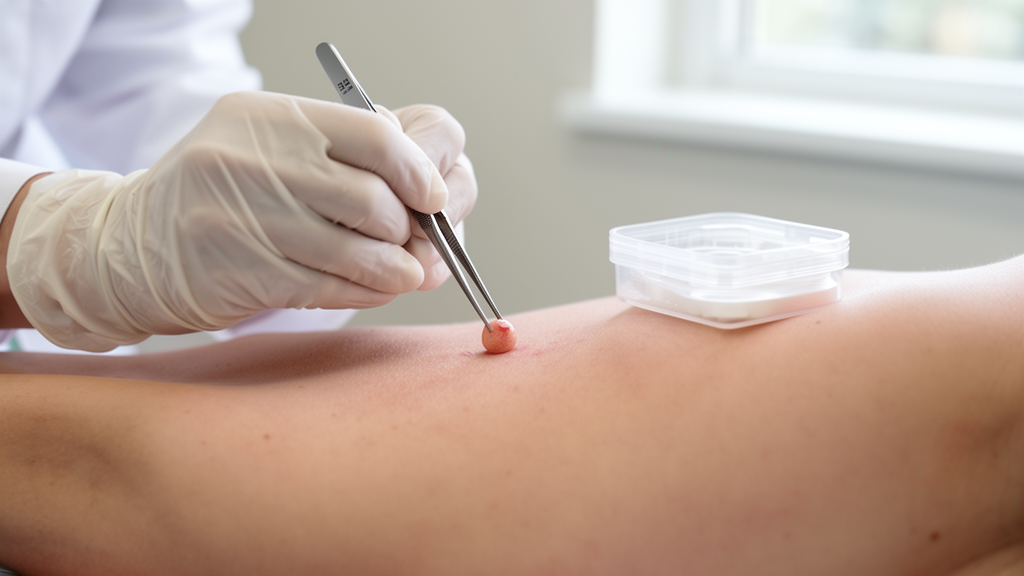
The central finding, that fibroblasts grown from a small skin biopsy can often supply the diagnostic information previously requiring muscle or liver tissue, rests on a biological insight that has matured over decades. Skin cells are remarkably cooperative in the laboratory: a punch biopsy about the size of a pencil eraser, taken under local anesthesia in an outpatient setting, yields cells that can be expanded indefinitely in culture dishes. Mitochondrial dysfunction is generally expressed throughout the body’s tissues, so fibroblasts frequently carry the same biochemical fingerprint as muscle or liver, the tissues traditionally sampled. Culturing the cells also offers an advantage that fresh tissue does not: the laboratory can grow enough material to repeat assays, run additional tests as new methods emerge, and bank samples for future reanalysis as knowledge advances.
For patients, the practical implications are substantial. A skin biopsy can be performed in a clinic visit with local anesthetic, avoiding the operating room, the general anesthetic, and the postoperative recovery that muscle and liver biopsies entail. General anesthesia itself carries particular risks for patients with mitochondrial disease, since metabolic stress during surgery can precipitate clinical deterioration, a concern that has long made invasive diagnostics a genuinely dangerous proposition for the sickest patients. Removing that barrier means that patients who previously might have deferred testing, or whose physicians hesitated to recommend surgery, can now pursue a diagnosis with far lower stakes. It also opens the door to testing at centers far from the few hospitals that perform specialized tissue biopsies, since a skin sample can be shipped to a reference laboratory.
The translation of research assays into routine clinical service is often the slowest step in medical progress, and the Colorado team has moved it forward in parallel with the science. Nearly all of the functional tests examined in the publication are now offered through the Mitochondrial Diagnostic Laboratory within the Department of Pathology and Laboratory Medicine at Children’s Colorado, which has grown into one of the largest providers of mitochondrial functional testing in North America, receiving samples from medical centers throughout the United States and Canada. This clinical infrastructure matters because it converts a published finding into something a physician anywhere can actually order, and because the volume of samples flowing through the laboratory feeds back into continuous refinement of the assays themselves.
The study also benefited from sustained public investment in rare disease research infrastructure. Support from the National Institutes of Health through the North American Mitochondrial Disease Consortium, a network established to link patients, clinicians, and investigators across institutions, made a study of this geographic scope feasible. Consortia of this kind solve a fundamental problem in rare disease research: no single center sees enough patients to power meaningful studies, but a coordinated network can. Funding from the Children’s Hospital Colorado Foundation and the University of Colorado Foundation supplemented the federal support, illustrating how philanthropy often bridges the gap for conditions that fall outside commercial incentives.
Looking ahead, the most promising model is likely to be complementary rather than competitive: genetic sequencing to identify candidate variants, followed by functional testing to confirm or exclude their significance. When a sequencing report lists several variants of uncertain significance, demonstrating that the patient’s cells actually show impaired respiratory chain function can tip the balance toward a diagnosis, while normal function in the relevant pathway can redirect the search. Conversely, a clear functional defect can guide which of the many uncertain variants deserves attention. This interplay between reading the genome and testing its output reflects a broader shift in genetics toward multidisciplinary interpretation, and for mitochondrial disease it may finally shorten journeys that families have too often measured in decades.
Subject of Research: Functional diagnostic testing of fibroblasts from skin biopsies for mitochondrial disease diagnosis
Article Title: International study validates less invasive approach to diagnosing mitochondrial disease
Article References: International study validates less invasive approach to diagnosing mitochondrial disease. (n.d.). Original publication
Image Credits: AI Generated
DOI: Not provided
Keywords: mitochondrial disease, skin biopsy, fibroblasts, functional testing, genetic testing, Children's Hospital Colorado, EMBO Molecular Medicine, rare disease, diagnosis, mitochondria, NAMDC, precision medicine
News Source: Ophelia Keating. (October 18, 2026). Skin Biopsy Outperforms Invasive Surgery in Global Mitochondrial Disease Study. Scienmag.