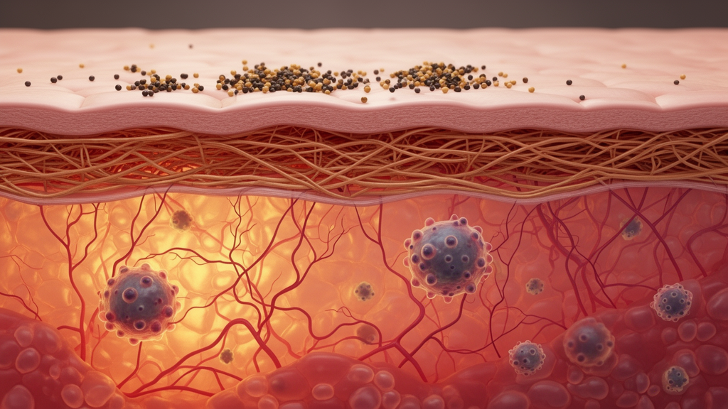
A prospective pilot study indicates that skin hyperpigmentation commonly classified as a post-inflammatory condition actually exhibits persistent dermal and interstitial inflammation long after primary skin lesions clinically resolve. The researchers propose redefining this condition as persistent and inflammatory hyperpigmentation, or PAIH, challenging the long-standing assumption that hyperpigmentation represents a static pigmentary residue left behind after cutaneous inflammation subsides. Conducted at the Johns Hopkins Cutaneous Translational Research Program, the investigation integrated non-invasive optical spectroscopy, objective colorimetry, structured participant questionnaires, and blinded histopathological analysis of paired tissue biopsies. By evaluating affected skin alongside adjacent unaffected control tissue in adult participants, the research team identified ongoing immune infiltration and epidermal remodeling beneath hyperpigmented macules resulting from common inflammatory skin conditions such as acne vulgaris, atopic dermatitis, and plaque psoriasis.
Post-inflammatory hyperpigmentation, commonly designated as PIH, represents an exceptionally frequent pigmentary alteration triggered by cutaneous injury, dermatological procedures, or inflammatory dermatoses. Although PIH can arise in all skin phenotypes, it occurs more frequently and presents with greater clinical prominence in individuals with skin of color, frequently causing substantial psychosocial distress and diminishing quality of life. Despite its extensive prevalence in outpatient practice, the biological mechanisms driving PIH pathogenesis and persistence have remained poorly defined. Conventional dermatological thinking assumes that tissue inflammation wanes as active inciting lesions clear, prompting clinical management to discontinue anti-inflammatory interventions and transition solely to pigment-lightening therapies. This prospective pilot trial was designed to examine whether microscopic inflammatory activity truly ceases after macroscopic lesions heal or whether persistent cellular signaling continues to drive ongoing melanogenesis within the dermis and epidermis.
The study enrolled twenty-three adult volunteers, including eighteen African American individuals, three Asian individuals, and two White individuals. Eligible participants were eighteen years of age or older, in good general health, and presented with hyperpigmentation resulting from a verified prior inflammatory event. Protocol criteria excluded candidates with active interfering skin diseases, bleeding disorders, local anesthetic allergies, keloidal scarring tendencies, current pregnancy, lactation, or recent treatments capable of influencing tissue evaluation. Among the enrolled cohort, acne was the inciting condition in 43.5 percent of subjects, representing ten individuals, while atopic dermatitis or psoriasis accounted for 39.1 percent, representing nine individuals, and external trauma or procedures caused 17.4 percent, representing four individuals. Racial stratification demonstrated distinct etiology patterns: acne predominated among African American participants, psoriasis and atopic dermatitis occurred most frequently in Asian participants, and external trauma appeared most often among White participants.
Participant surveys detailed medical histories, lesion duration, anatomical distributions, and personal perceptions of skin changes. Duration assessments revealed that twelve subjects had experienced hyperpigmentation for more than three years, five subjects had persistent lesions lasting one to three years, three had lesions for one year, and three had lesions for six months, while no participants presented with durations under six months. Lesion duration showed no statistically significant association with the primary inciting dermatosis. Anatomically, forty-three hyperpigmented sites occurred on non-photoexposed regions such as the chest, back, abdomen, buttocks, and legs, whereas twenty-six lesions were located on photoexposed sites including the face, neck, and arms. Participants with hyperpigmentation persisting beyond three years demonstrated a statistically significant higher likelihood of reporting a positive family history of the condition and were significantly more inclined to seek professional medical care.
Questionnaire responses also highlighted notable demographic patterns in familial occurrence and treatment-seeking behavior. Male participants were significantly more likely than females to report a family history of hyperpigmentation, with 64 percent of males reporting familial occurrence compared to 22 percent of females. Furthermore, healthcare-seeking actions differed significantly by racial and ethnic background: African American participants were the most likely to seek clinical treatment, followed sequentially by Asian participants and White participants. Patients who consulted physicians for treatment also expressed significantly greater levels of dissatisfaction regarding their condition. The authors noted that in previous dermatological literature, women with skin of color prioritized resolving hyperpigmentation following acne at a rate of 41.6 percent compared to only 8.4 percent among White women, underscoring the acute psychosocial burden and clinical urgency of pigmentary sequelae across diverse patient groups.
To establish objective physical parameters of cutaneous pigmentation, investigators utilized standardized colorimetry alongside spectrophotometric evaluation. A Konica Nikon CR-400 colorimeter quantified skin brightness along the L* coordinate axis, where zero represents total black and one hundred represents pure white. Measurements confirmed that hyperpigmented macules exhibited significantly lower L* values than adjacent normal skin across all subjects, providing objective proof of relative darkening. Although the luminance difference, termed delta L*, showed no significant variance based on inciting condition or participant gender, it varied significantly across racial and ethnic groups. African American participants exhibited the smallest delta L* value at 4.26 plus or minus 3.79, Asian participants demonstrated an intermediate value of 8.24 plus or minus 2.82, and White participants presented the largest contrast between affected and normal skin at 9.71 plus or minus 2.09.
Cutaneous chromophores were evaluated using an ASD LabSpec 5000 spectrophotometer recording spectral reflectance across wavelengths from 350 to 1830 nanometers to determine absolute and relative melanin levels. Absolute melanin values in hyperpigmented lesions were highest among African American participants at 587.8 plus or minus 52.2, followed by Asian participants at 494.0 plus or minus 43.7, and White participants at 470.7 plus or minus 1.5. A linear mixed-effects model incorporating cubic splines with six knots confirmed that spectral reflectance curves showed no significant differences across distinct inciting causes of hyperpigmentation. Furthermore, relative melanin values did not differ significantly by primary disease etiology or racial background, nor did they correlate with whether individuals sought clinical treatment. These spectroscopic data indicate that while absolute melanin concentrations reflect baseline constitutive pigmentation, relative optical changes induced by hyperpigmentation behave consistently regardless of the primary disease trigger.
Blinded histopathological examination of paired cutaneous punch biopsies provided direct microscopic evidence supporting the researchers’ findings. Dermatopathologists evaluated twenty-seven hyperpigmented tissue specimens taken from one or two four-millimeter punch biopsies per participant alongside control biopsies from adjacent unaffected skin. Semi-quantitative microscopic scoring revealed that hyperpigmented skin exhibited a seventy-four-fold increase in epidermal acanthosis, a forty-four-fold increase in hyperkeratosis, and a forty-four-fold increase in rete ridge elongation compared to normal skin. Dermal structural changes were similarly prominent, including a 17.5-fold elevation in dermal fibrosis, a nineteen-fold increase in plasma cells, and a 1.5-fold increase in dermal melanophages trending toward statistical significance. The marked epidermal thickening and architectural elongation observed across biopsy sections indicate that structural cellular expansion in the epidermis actively reinforces the visual perception of skin darkening in hyperpigmented lesions.
Remarkably, microscopic evaluation demonstrated ongoing inflammatory cell infiltration within hyperpigmented tissue long after the clinical resolution of active dermatitis, acne, or psoriasis plaques. Biopsy analysis documented a 16.5-fold increase in mid-dermal inflammation and a twenty-two-fold increase in interstitial inflammation relative to unaffected control tissue. Immunohistochemical staining confirmed a 1.7-fold elevation in CD3-positive T lymphocytes and a 6.1-fold increase in CD20-positive B lymphocytes, with CD3-positive T cells constituting the predominant infiltrate in perivascular and interstitial spaces. Fontana-Masson silver staining and hematoxylin and eosin imaging demonstrated dense melanin particle clustering near the dermal-epidermal junction and superficial blood vessels. Melanin particles and staining intensity were elevated in the papillary dermis of both acne and psoriasis samples, with acanthosis presenting in a thicker, more uniform pattern in psoriasis-associated hyperpigmentation than in acne-associated tissue.
Based on these optical and cellular observations, the investigators argue that classifying this pathology as post-inflammatory represents an inaccurate description that mischaracterizes the underlying disease process. Because interstitial and dermal inflammation actively persists, the authors propose renaming the entity persistent and inflammatory hyperpigmentation to reflect ongoing immunological engagement. In acne-induced hyperpigmentation, prior studies demonstrated subclinical inflammation inside comedones lacking external redness, where arachidonic acid cascades release prostaglandins and leukotrienes that stimulate melanosome transfer to keratinocytes. In psoriasis, persistent dermal immune cascades involve tumor necrosis factor and interleukin-17 pathways that modulate melanocyte dendricity and proliferation, while in atopic dermatitis models, ongoing signaling involving mast cells, interleukin-33, and histamine promotes sustained melanogenesis. Framing hyperpigmentation as a persistent inflammatory disorder provides a sound biological rationale for sustaining anti-inflammatory therapy rather than relying exclusively on post-flare cosmetic pigment clearance.
These findings indicate that therapeutic management may require prolonged anti-inflammatory regimens even after inciting conditions appear clinically resolved, combined with pigment-reducing agents such as topical hydroquinone, retinoids, keratolytics, and antioxidants. Because acanthosis contributes substantially to clinical darkening, keratolytics and retinoids may provide structural benefits by normalizing epidermal thickness. Comprehensive photoprotection covering ultraviolet B, long-wave ultraviolet A, and visible light remains foundational, particularly for skin of color. The authors noted key study limitations, including a pilot sample of twenty-three participants at a single institution, lack of long-term follow-up, recall bias in surveys, and absence of Fitzpatrick phototyping or sunlight exposure records. Predominantly enrolling African American subjects with acne, psoriasis, or atopic dermatitis also limits immediate generalizability. The researchers concluded that larger multicenter prospective trials are required to confirm these histopathological changes and evaluate prolonged anti-inflammatory combination therapies.
Subject of Research: Persistent dermal inflammation and histopathological alterations in post-inflammatory hyperpigmentation
Article Title: Rethinking post inflammatory hyperpigmentation: A prospective pilot study supporting a persistent not post-inflammatory process
Article References: Elbuluk, N., Mu, E. W., Hurtado, A. C. M., Syder, N. C., Erdag, G., Gomez-Ospina, N., Colantuoni, E., Taube, J. M., Okoye, G. A., & Kang, S. (2026). Rethinking post inflammatory hyperpigmentation: A prospective pilot study supporting a persistent not post-inflammatory process. Archives of Dermatological Research, 318(1), Article 559. https://doi.org/10.1007/s00403-026-05017-4
Image Credits: AI Generated
DOI: 10.1007/s00403-026-05017-4
Keywords: Post-Inflammatory Hyperpigmentation, Persistent and Inflammatory Hyperpigmentation, Skin of Color, Cutaneous Histopathology, Dermal Inflammation, Interstitial Inflammation, Epidermal Acanthosis, CD3 Positive Lymphocytes, CD20 Positive Lymphocytes, Colorimetry, Reflectance Spectrophotometry, Acne Vulgaris, Atopic Dermatitis, Plaque Psoriasis, Melanogenesis, Dermal Fibrosis, Melanophages
News Source: Ophelia Keating. (November 12, 2026). Skin Biopsies Reveal Ongoing Tissue Inflammation Beneath Resolved Post-Inflammatory Hyperpigmentation. Scienmag.