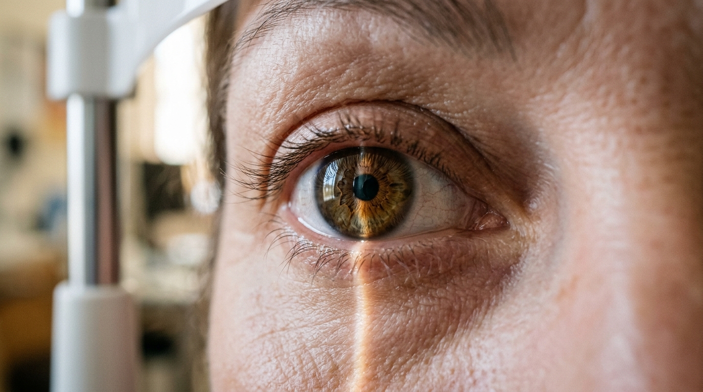
A single topical instillation of low-concentration atropine across a range of dosing strengths produces no measurable change in axial length or retinal and choroidal thickness over twenty-four hours in healthy young adults, according to a randomized, double-masked, repeated-measures investigation published in the journal Eye and Vision. The study, conducted by researchers Barsha Lal and Lisa A. Ostrin, systematically evaluated how healthy human eyes respond in the acute window following administration of topical atropine drops ranging from 0.01 percent to 0.1 percent. Although the researchers observed that ocular axial dimensions and structural tissue layers remained stable throughout the testing period, high-resolution vascular imaging revealed a transient, time-dependent reduction in superficial retinal microvascular perfusion density one hour after drop delivery. These physiological findings clarify the acute ocular behavior of muscarinic receptor antagonism and demonstrate that the structural changes frequently reported during long-term clinical myopia management do not emerge following an isolated single treatment session.
The global public health context surrounding myopia progression underscores why researchers are closely scrutinizing the exact biological actions of atropine. Current international projections estimate that myopia affects approximately 34 percent of the global population and will rise to 50 percent by the year 2050, with uncorrectable visual impairment in the United States projected to be between 27 percent and 43 percent attributable to myopia by mid-century. Because individuals with myopia face elevated lifetime risks for sight-threatening ocular pathologies, delaying onset and slowing refractive elongation carry substantial societal and clinical benefits. Although optical, environmental, behavioral, and surgical interventions exist, topical atropine represents the most widely prescribed pharmacological therapy proven to slow childhood myopia progression across clinical trials worldwide. Atropine acts as a non-specific muscarinic antagonist that blocks all five known acetylcholine muscarinic receptor subtypes, designated M1 through M5. These receptors are widely distributed throughout ocular structures, including the iris, ciliary body, lens, retina, and sclera, with animal studies providing evidence of muscarinic receptors within the vascular choroid.
Historically, atropine was introduced for childhood refractive control based on its cycloplegic properties, which temporarily paralyze the ciliary muscle and inhibit accommodation under the early hypothesis that excessive visual accommodation drove axial elongation. Subsequent experimental evidence from avian models, however, demonstrated that atropine prevented experimental myopia through a non-accommodative mechanism, establishing that its precise biological pathways remain incompletely understood. Muscarinic blockade has been shown to modulate biochemical signaling and alter scleral extracellular matrix remodeling, which may contribute to anti-myopia efficacy. Early human trials relied on strong 1.0 percent atropine solutions, which demonstrated robust efficacy in slowing progression and axial elongation, but clinical utility was frequently compromised by severe adverse side effects, including pupil dilation, photophobia, glare, and blurred near vision. Consequently, modern practice has shifted toward low-concentration formulations at or below 0.1 percent, where meta-analyses show dose-dependent efficacy balanced against acceptable tolerability profiles, making detailed structural and microvascular evaluations at lower concentrations essential.
To investigate whether acute structural changes explain atropine action, the Houston-based research team designed a double-masked, randomized, repeated-measures trial involving healthy adult volunteers aged 18 to 35 years. The experimental cohort comprised twenty participants who completed the full multi-visit regimen, consisting of ten females and ten males with a mean age of 25.5 years, a standard deviation of 3.4 years, and an age range spanning 19 to 33 years. Enrolled subjects presented with a mean spherical equivalent refraction in the evaluated right eye of minus 1.91 diopters, with a standard deviation of 2.24 diopters and an overall range from minus 6.62 to plus 0.75 diopters. The cohort encompassed twelve myopic participants, six emmetropic participants, and two hyperopic participants. One additional enrolled subject withdrew prior to study completion after experiencing prolonged pupil dilation that persisted for more than one month following their initial session, which subsequent experimental unmasking confirmed had involved the highest tested dose of 0.1 percent atropine.
The study protocol systematically evaluated five separate experimental conditions for each participant, comprising four compounded ophthalmic atropine concentrations of 0.01 percent, 0.025 percent, 0.05 percent, and 0.1 percent, alongside an identical placebo formulation containing 0.0 percent active drug. The sterile solutions were formulated locally with a hypromellose vehicle, phosphate buffer, ethylene diamine tetraacetic acid, and sterile water for irrigation, set to a pH of 4.5 for active atropine and 5.0 for the placebo. The five sessions were administered in randomized order across visits spaced one to three weeks apart to prevent carryover influences. Experimental visits were scheduled strictly between 8:30 a.m. and 11:30 a.m. to minimize confounding effects from normal diurnal fluctuations, with each participant beginning every personal session within a consistent one-hour window. Participants avoided contact lenses, alcohol, caffeine, tobacco, and vigorous exercise on testing days, and relaxed while watching distant television at six meters for ten minutes prior to recordings to stabilize baseline physiological ocular parameters.
Ocular dimensions and vascular architectures were quantified across three discrete time points during every visit: immediately prior to instillation at baseline, at one hour post-instillation, and at twenty-four hours post-instillation. Optical biometry performed with a MYAH device captured ocular axial length, averaging three repeated scans per session. Retinal and choroidal structures were recorded using high-resolution spectral-domain optical coherence tomography radial scans centered on the fovea, analyzed with custom MATLAB algorithms to delineate boundaries at the internal limiting membrane, retinal pigment epithelium, and choroid-sclera interface across both the central 1.0-millimeter diameter and an outer 1.0 to 3.0-millimeter annulus. Concurrently, optical coherence tomography angiography mapped microvascular perfusion across the superficial vascular complex, deep vascular complex, and choriocapillaris within the central 1.0-millimeter zone and an outer 1.0 to 2.5-millimeter annulus, alongside foveal avascular zone area, after applying individualized lateral magnification scaling and projection artifact removal.
Analysis of structural outcomes using two-way repeated-measures analysis of variance revealed that neither axial length nor retinal or choroidal thickness underwent significant alterations across the study timeline. Specifically, no statistically significant changes were observed in axial length, central 1.0-millimeter retinal thickness, outer annular retinal thickness, central choroidal thickness, or annular choroidal thickness across any tested drug concentration or follow-up time point, yielding p-values exceeding 0.05 throughout. Furthermore, the statistical modeling detected no significant interaction between drug concentration and measurement timing. Instrumental test-retest reliability demonstrated high precision, exhibiting a mean baseline measurement difference of minus 0.1 micrometers for central retinal thickness and 1.3 micrometers for central choroidal thickness between repeated baseline evaluations. These stable findings confirm that a single low-dose atropine administration does not trigger rapid mechanical thinning, fluid shifting, or acute tissue expansion within the posterior ocular coat over twenty-four hours in healthy adult eyes.
In contrast to the absence of bulk tissue dimensional shifts, optical coherence tomography angiography identified distinct microvascular dynamics confined to the superficial retinal vasculature. In the annular region spanning 1.0 to 2.5 millimeters from the fovea, perfusion density within the superficial vascular complex exhibited a statistically significant decrease over time following drop instillation, registering an overall analysis of variance p-value of 0.02. This vascular shift was purely time-dependent rather than concentration-dependent, as variation across drug concentrations showed no statistically significant effect, with p-values exceeding 0.05 and no significant interaction observed between concentration and time. Bonferroni-adjusted post hoc pairwise evaluations revealed that superficial vascular complex perfusion density declined significantly from baseline at the one-hour mark by an average of minus 1.3 percent, with a standard deviation of 1.2 percent and a p-value of 0.03. By twenty-four hours, this perfusion suppression largely resolved, showing a non-significant mean difference of minus 0.4 percent, with a standard deviation of 0.6 percent and a p-value of 0.28.
Microvascular evaluations across deeper vascular beds and alternative macular zones demonstrated stability following treatment. Perfusion density in the central 1.0-millimeter foveal diameter showed no significant changes over time or across drug concentrations for either the superficial or deep vascular complexes. Similarly, within the outer 1.0 to 2.5-millimeter annulus, perfusion density across the deep vascular complex and the underlying choriocapillaris showed no statistically significant alterations, maintaining p-values greater than 0.05. The total area of the foveal avascular zone likewise remained stable throughout the post-instillation periods. The investigators noted that the localized microvascular response observed within the superficial annulus may reflect the greater concentration of medium and larger arterioles and venules located within this specific retinal layer, whereas the deeper plexuses and choriocapillaris consist primarily of fine capillary meshes. Although concentration effects did not achieve statistical significance, the greatest absolute numerical drop in superficial perfusion and the highest absolute numerical increase in choriocapillaris perfusion occurred under the 0.1 percent atropine condition.
The lack of acute choroidal thickening in this trial provides an informative contrast to previously published literature and highlights differences between acute isolated exposures and chronic therapeutic regimens. Prior pediatric and adult investigations have reported conflicting choroidal responses, ranging from subfoveal thickening within one hour or one week to absence of change, or even choroidal thinning following six months of continuous daily therapy. Because previously published pupil and accommodative data from this exact trial cohort established clear dose-dependent pupil dilation and accommodative paresis at one and twenty-four hours, the absence of posterior structural shifts cannot be attributed to inadequate ocular penetration. The authors noted several operational boundaries to their study, including a modest sample size of twenty young adults, an acute single-dose administration schedule that does not capture cumulative daily exposure, and the intentional exclusion of high myopes. They concluded that while acute low-dose atropine does not alter axial length or posterior segment thickness over twenty-four hours, it induces a transient, time-dependent reduction in superficial retinal perfusion that warrants continued physiological tracking across clinical and research settings.
Subject of Research: Short-term ocular structural and microvascular effects of low-concentration atropine eye drops in healthy young adults
Article Title: Short-term effects of atropine on the retina and choroid in young adults
Article References: Lal, B., & Ostrin, L. A. (2026). Short-term effects of atropine on the retina and choroid in young adults. Eye and Vision, 13(1), Article 8. https://doi.org/10.1186/s40662-026-00477-1
Image Credits: AI Generated
DOI: 10.1186/s40662-026-00477-1
Keywords: atropine, myopia control, choroid, retina, axial length, optical coherence tomography, optical coherence tomography angiography, superficial vascular complex, choriocapillaris, deep vascular complex, foveal avascular zone, ophthalmology, ocular physiology, muscarinic antagonists, refractive error, eye drops, ocular microvasculature
News Source: Ophelia Keating. (November 11, 2026). Single Low-Dose Atropine Drops Do Not Alter Retinal Thickness in Adults. Scienmag.