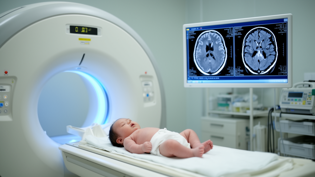
In a retrospective cohort study of 109 neonates treated at Hunan Children’s Hospital in China, the anatomical distribution of intracranial bleeding was associated with the severity of imaging-defined post-hemorrhagic ventricular dilatation, but hemorrhage distribution was not independently associated with mortality or neurodevelopmental abnormality. Infants with combined intraventricular and cerebral parenchymal hemorrhage had the highest prevalence of moderate-to-severe ventricular dilatation, followed by those with isolated intraventricular hemorrhage; infants with isolated parenchymal hemorrhage had the lowest prevalence.
The study included neonates hospitalized with intraventricular hemorrhage, cerebral parenchymal hemorrhage, or both between 2015 and 2018. Of the 109 participants, 44, or 40.4%, had isolated intraventricular hemorrhage, 41, or 37.6%, had isolated cerebral parenchymal hemorrhage, and 24, or 22.0%, had combined intraventricular and parenchymal involvement. Moderate-to-severe imaging-defined post-hemorrhagic ventricular dilatation occurred in 61.4% of the isolated intraventricular hemorrhage group, 24.4% of the isolated parenchymal hemorrhage group, and 70.8% of the combined group. The severity differences among these groups were statistically significant, with a reported P value below 0.001, indicating an association between ventricular involvement and more severe ventricular enlargement in this cohort.
Clinical outcomes did not differ significantly among the three hemorrhage groups. Across the cohort, 41 neonates died, representing 37.6% of those studied; 21, or 19.3%, had a recorded neurodevelopmental abnormality; and 47, or 43.1%, had favorable outcomes. The overall distribution of outcomes among the hemorrhage groups was not significant, with a reported P value of 0.693. Multivariable logistic regression identified lower gestational age and higher clinical severity scores as factors independently associated with mortality. Thus, while hemorrhage distribution corresponded with the degree of ventricular dilatation, the analysis did not identify it as an independent correlate of death or neurodevelopmental abnormality.
The investigators also evaluated cerebrospinal fluid drainage and neurosurgical intervention. Either form of treatment was associated with lower odds of neurodevelopmental abnormality, with an odds ratio of 0.115, a 95% confidence interval of 0.022–0.595, and a P value of 0.010. This association cannot be interpreted as evidence that drainage or surgery prevented neurological impairment. The retrospective, single-center, nonrandomized design allowed selection and survivorship bias to influence which infants received particular interventions and which outcomes were observed, preventing causal or preventive-effect inferences. The confidence interval was also wide, reflecting uncertainty in the estimated association.
Brain imaging supplied complementary anatomical information for the assessment. Magnetic resonance imaging, performed in a subset of infants, additionally identified deep, posterior-fossa, subarachnoid, and small focal hemorrhagic lesions. The authors characterize ultrasound and MRI as complementary for evaluating neonatal brain injury and ventricular enlargement. Overall, the findings indicate that hemorrhage involving the ventricles identifies infants with a greater observed burden of significant ventricular dilatation, while gestational maturity and systemic disease burden remain important determinants of outcome. The evidence does not establish whether hemorrhage distribution independently affects survival or neurological development, nor whether cerebrospinal fluid-directed treatment improves those outcomes.
Subject of Research: Associations among hemorrhage distribution, post-hemorrhagic ventricular dilatation, and outcomes in neonates with intraventricular or cerebral parenchymal hemorrhage.
Article Title: Hemorrhage distribution, imaging-defined post-hemorrhagic ventricular dilatation, and clinical outcomes in neonates with intraventricular and cerebral parenchymal hemorrhage: a single-center retrospective cohort study
Article References: Zhang, X., Cai, Y., Huang, S., Shi, H., Chen, L., Wu, S., & Xiang, Y. (2026). Hemorrhage distribution, imaging-defined post-hemorrhagic ventricular dilatation, and clinical outcomes in neonates with intraventricular and cerebral parenchymal hemorrhage: a single-center retrospective cohort study. Frontiers in Pediatrics, 14, Article 1965213. https://doi.org/10.3389/fped.2026.1965213
Image Credits: AI Generated
DOI: 10.3389/fped.2026.1965213
Keywords: neonatal brain hemorrhage, intraventricular hemorrhage, cerebral parenchymal hemorrhage, post-hemorrhagic ventricular dilatation, neonatal mortality, neurodevelopment, cranial ultrasound, brain MRI, Hemorrhage, distribution, imaging-defined, post-hemorrhagic
News Source: Harold Sullivan. (October 22, 2026). Single-center cohort links ventricular bleeding to greater neonatal ventricular dilation. Scienmag.