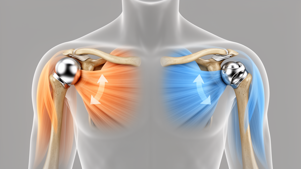
Reverse total shoulder replacement has been quietly taking over operating rooms around the world. International joint registry data show that the procedure has nearly doubled over the past decade and now accounts for a larger and growing share of shoulder replacement surgeries than the anatomic design it originally supplemented. That shift has raised an uncomfortable question for surgeons: when a patient has osteoarthritis but a fully functioning rotator cuff, which implant should go in?
Researchers at the University of Rostock used a validated musculoskeletal multibody simulation framework, known as the Rostock Shoulder Software-in-Loop framework, to pit the two implant designs against each other under strictly identical conditions. The model, built in the SIMPACK multibody environment, represented the upper extremity as a kinematic chain of seven rigid bone segments reconstructed from computed tomography data of the Visible Human Male dataset, a 58-kilogram male subject. Forty-eight muscles, discretized into 152 force elements, drove the motion. Crucially, the glenohumeral joint was not simplified into the ball-and-socket abstraction used in many earlier models. Instead, it carried six degrees of freedom with explicit, frictional contact between the actual articulating implant surfaces, allowing the team to track joint translations, contact pressures, and the wandering center of pressure that simpler models cannot capture.
The virtual surgery paired two commercially available systems from the same manufacturer. The anatomic configuration used the Affinis system with a polyethylene glenoid component and a cobalt-chromium-molybdenum humeral head, while the reverse configuration used the Affinis Inverse with a polyethylene glenosphere and a ceramic humeral inlay. Both were virtually implanted according to the manufacturer’s surgical technique and verified by shoulder surgeons. The simulated task was a 120-degree thoracohumeral arm elevation in the scapular plane. The rotator cuff was modeled as fully intact and functional in both designs, a deliberate choice meant to represent the select group of older patients with primary osteoarthritis in whom either implant might be considered, and to isolate the pure effect of implant geometry from the confounding influence of cuff deficiency.
The headline result concerns muscle strategy. Switching from the anatomic to the reverse design cut the mean middle deltoid force by 16 percent, with the peak dropping from 42.16 percent of body weight, about 240 newtons, to 31.09 percent of body weight, roughly 177 newtons. The reverse design achieves this by medializing and inferiorizing the joint’s center of rotation by approximately 27 millimeters, which lengthens the deltoid’s lever arm and increases its mechanical advantage for lifting the arm. Forces in the anterior and posterior deltoid barely changed, but every rotator cuff muscle worked less hard in the reverse configuration. The maximum forces of the infraspinatus, subscapularis, and supraspinatus all fell substantially, and the supraspinatus muscle ran consistently shorter along its altered path throughout the elevation.
To quantify this shift, the team introduced a deltoid-to-cuff ratio, the summed deltoid forces divided by the summed rotator cuff forces. In the anatomic design, the ratio started near 0.95, meaning the cuff carried slightly more load, and stayed close to balance until a crossover at around 58 degrees of elevation, after which the deltoid took over. In the reverse design, the ratio never dropped below one. It began at 1.76, climbed to 2.20 at 90 degrees, and peaked at 3.04 at the top of the motion. In other words, the reverse implant performs arm elevation through a deltoid-driven solution across the entire range, relying far less on the cuff force couples that stabilize the anatomic joint.
The joint loading picture was equally striking. Compressive contact forces across the implant surfaces fell by 26.5 percent in the reverse design, and the total resultant joint force fell by 33.4 percent. Contact pressure told the same story from a different angle: the anatomic glenoid component saw pressures rising to a peak of 4.12 megapascals, while the reverse glenosphere stayed between 0.6 and 1.17 megapascals, roughly 3.5 times lower. The reason is geometry. The conforming reverse articulation spread the same, smaller load over a contact area nearly three times larger, peaking at 343 square millimeters compared with 124 square millimeters for the anatomic design. Lower compression and pressure, the authors note, may reduce mechanical loading on the fixation interface and the risk of glenoid loosening.
But the reverse design carried a trade-off, and it showed up in shear. Superior shear force in the reverse joint rose earlier during elevation and peaked at 20.09 percent of body weight at 50 degrees, 98.7 percent higher than the anatomic peak of 10.11 percent of body weight, which arrived later at 75 degrees. Expressed as a shear-to-compression instability ratio, the reverse design averaged 0.39, compared with 0.12 for the anatomic design, a nearly threefold difference driven overwhelmingly by the superior-inferior direction. The authors are careful about interpretation: this force-based metric does not account for the reverse implant’s geometric conformity and semi-constraint, so a higher ratio does not directly translate into higher clinical dislocation risk. It signals a less compression-dominant loading balance, particularly in the early phase of elevation, which corresponds to the arm position clinicians already consider most vulnerable in reverse shoulders.
The center of pressure trajectories diverged in ways that matter for implant longevity. In the anatomic design, the contact point stayed posterior and mostly superior across most of the motion, a pattern the authors link to potential posterior edge loading and component loosening. In the reverse design, the contact point migrated dramatically in the superior-inferior direction, shifting from an inferior starting position of minus 10.8 millimeters superiorly, with maximum displacement of 15.48 millimeters versus 7.32 millimeters for the anatomic design. Eccentric contact creates rocking moments at the implant-bone interface, a known contributor to loosening, and the contact area and pressure patterns generated here can feed directly into future polyethylene wear models. Notably, prior simulations have estimated up to five times higher volumetric wear rates in reverse designs precisely because their larger contact area trades lower linear wear for higher volumetric wear.
Joint translations revealed the two designs’ fundamentally different stabilizing philosophies. The anatomic implant allowed a relatively mobile humeral head, with its largest translations in the superior-inferior direction, up to nearly a millimeter, and persistent posterior positioning. The reverse implant constrained the humeral inlay to a tightly guided path around the glenosphere, with negligible posterior-anterior displacement of just 0.05 millimeters and total translation amplitudes below half a millimeter, reflecting its deep, conforming socket. The joint, in effect, moves the same way through two entirely different mechanical strategies: one trusting muscle balance, the other trusting geometry.
The authors are candid about the study’s limits. The simulation used a single generic male anatomy rather than a patient cohort, one implant system from one manufacturer in one positioning, a single prescribed elevation motion, and no explicit capsular or ligamentous restraints. Muscle force predictions also depend on the assumed recruitment strategy, so absolute values should be read cautiously even if the comparative differences align with prior experimental and computational work. The findings apply specifically to the intact-cuff condition and cannot be extrapolated to cuff-tear arthropathy or revision surgery, where cuff integrity substantially reshapes joint loading. Still, within that scope, the study delivers a rare controlled, side-by-side quantification of what surgeons trade when they choose a reverse implant for a patient whose rotator cuff still works: less deltoid effort and less joint compression, but higher superior shear, a less favorable shear-to-compression balance, and a different pattern of contact migration across the implant surface.
Subject of Research: A computational comparison of glenohumeral joint biomechanics between anatomic and reverse total shoulder replacement under intact rotator cuff conditions.
Article Title: Anatomic versus reverse total shoulder replacement with an intact rotator cuff: a comparative musculoskeletal simulation study of glenohumeral joint biomechanics
Article References: Soodmand, I., Tischer, T., Herrmann, S., Klose, T., Ruehrmund, L., Lutter, C., Woernle, C., Bader, R., & Kebbach, M. (2026). Anatomic versus reverse total shoulder replacement with an intact rotator cuff: a comparative musculoskeletal simulation study of glenohumeral joint biomechanics. Arthroplasty, 8(1), Article 72. https://doi.org/10.1186/s42836-026-00432-4
Image Credits: AI Generated
DOI: 10.1186/s42836-026-00432-4
Keywords: total shoulder replacement, reverse shoulder arthroplasty, anatomic shoulder replacement, rotator cuff, glenohumeral joint, biomechanics, musculoskeletal simulation, multibody modeling, joint contact forces, osteoarthritis, deltoid muscle, implant design
News Source: Ophelia Keating. (October 29, 2026). Simulation study maps how reverse and anatomic shoulder implants load the joint differently. Scienmag.