Medical physics researchers evaluating metal artifact mitigation in image-guided procedures have demonstrated that adding a dedicated zero-point-five millimeter silver filter to an interventional multidetector computed tomography scanner substantially suppresses needle-induced artifacts across an anthropomorphic chest phantom. By hardening the diagnostic x-ray beam prior to patient entry, the additional filtration diminished severe photon starvation and localized beam-hardening distortions generated along an eighteen-gauge biopsy trocar without degrading baseline background image noise. The experimental assessment, performed across comparable radiation dose levels fixed at one milligray volume computed tomography dose index, revealed that the filtered beam energy noticeably restricted peripheral streak propagation, reduced core needle blooming, and mitigated hypodense shadowing at the instrument tip. Because interventional computed tomography demands rapid, intraprocedural visualization to verify trajectory corrections in real time, standard post-processing metal artifact reduction algorithms are rarely deployed due to prohibitive computational reconstruction latency. The physical filtration strategy examined in this phantom investigation addresses this operational bottleneck by attenuating artifact generation at the source of emission, yielding clearer anatomical margins surrounding metallic instruments during simulated percutaneous tissue sampling.
Percutaneous interventional radiology procedures, including core biopsies and targeted thermal tumor ablations, depend fundamentally on the clear visual delineation of target lesions and adjacent critical neurovascular structures. During these delicate interventions, clinicians advance rigid metallic trocars, introducer cannulas, and microwave ablation antennas directly through soft parenchymal tissues under intermittent computed tomography guidance. However, the introduction of dense metal objects into the scanning field introduces severe physical degradation to the resulting cross-sectional image slices. Prior clinical observations cited in the evaluation highlight that the proportion of insufficiently visualized anatomical targets increases dramatically from ten point five percent on pre-biopsy scans to forty-four point seven percent once the metal introducer needle enters the targeted lesion. Techniques such as microwave ablation similarly require millimeter-level spatial precision between the active applicator antenna and target margins, emphasizing the acute need for effective intraprocedural artifact suppression strategies that do not hinder operational clinical workflow.
The physical mechanisms governing metal artifact creation in computed tomography stem from fundamental interactions between ionizing photons and dense metallic elements. This preferential absorption selectively hardens the transmitted x-ray spectrum, producing characteristic beam-hardening errors that manifest as spurious dark bands and shading across reconstruction planes. Concurrently, the extreme linear attenuation coefficient of dense metals causes acute photon starvation along trajectory projections passing through the longest axis of the instrument, depriving detector elements of adequate photon counts and inducing severe, high-amplitude streak artifacts punctuated by false hypointense and hyperintense bands. Secondary physical effects, including scattered radiation, nonlinear partial volume averaging, and magnified image noise within corrupted sinogram channels, further deteriorate visualization. While software-based iterative metal artifact reduction algorithms have been engineered for fixed surgical implants such as orthopedic hip prostheses, their sinogram interpolation methods risk deleting genuine anatomical structures and often generate secondary peripheral blooming artifacts, while remaining absent from real-time interventional fluoroscopy suites.
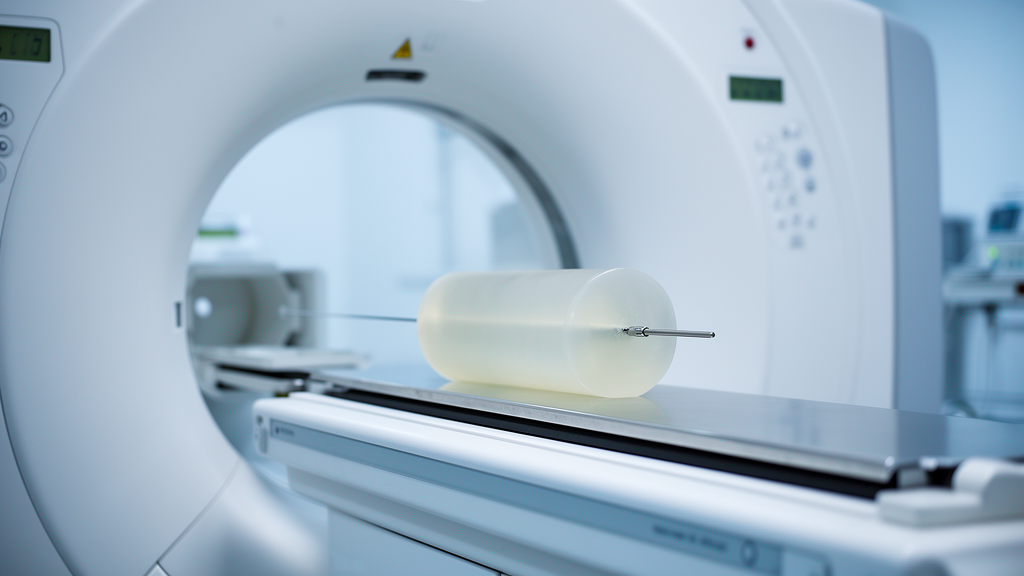
To evaluate whether pre-patient hardware beam filtration could mitigate these destructive distortions without computational latency, the investigative team conducted controlled phantom acquisitions utilizing a commercial multidetector computed tomography system. An anthropomorphic torso phantom, specifically the Lungman model from Kyoto Kagaku designed to simulate human thoracic anatomy, was configured with an eighteen-gauge, fifteen-centimeter Chiba biopsy needle and its surrounding coaxial sheath aligned horizontally within the lung parenchyma. The phantom was scanned using an Aquilion One Prism Edition scanner manufactured by Canon Medical USA, which incorporates a specialized, switchable zero-point-five millimeter silver filter designated SilverBeam. In standard diagnostic protocols, this silver filter increases the effective beam energy at one hundred and twenty kilovolts from sixty-six point two kiloelectron volts to eighty point nine kiloelectron volts, representing a twenty-two percent elevation in spectral hardness that closely mirrors the beam-hardening performance documented with tin filtration. The research protocol systematically compared this silver-filtered one hundred and twenty kilovolt configuration against three conventional unfiltered beam energies operated at one hundred, one hundred and twenty, and one hundred and thirty-five kilovolts.
Standardizing the experimental conditions across all scanning acquisitions was essential for validating comparative image quality and artifact severity. For every acquisition sequence, the volume computed tomography dose index was rigorously maintained at a constant one milligray, matching the conservative low-dose thresholds typically applied during routine interventional lung procedures to minimize cumulative radiation exposure to patients and operators. Automated tube current modulation was deliberately disabled to eliminate variable photon flux adjustments during rotational acquisitions. All volumetric datasets were subsequently reconstructed utilizing a standard filtered back projection algorithm paired with an FC18 reconstruction kernel, generating axial slices with a slice thickness of two millimeters and an image reconstruction matrix of five hundred and twelve by five hundred and twelve pixels. By maintaining identical scanner geometry, reconstruction mathematics, and radiation dose constraints across every acquisition, the investigators ensured that any observed modifications in artifact severity, spatial resolution, or background tissue noise could be directly attributed to the spectral alterations introduced by the silver filter.
Quantifying the spatial extent and amplitude of artifact distortions required a systematic profiling protocol established across three distinct spatial zones surrounding the inserted biopsy needle. High-resolution profiles were mapped perpendicular to the needle shaft to assess core blooming, across the hypointense region immediately anterior to the instrument tip to measure needle tip shadowing, and throughout the distal streak field to evaluate forward trajectory degradation. A fourth longitudinal profile tracked artifact decay along the forward trajectory vector to establish total artifact range until Hounsfield unit values returned to baseline background tissue levels. To achieve precise spatial sampling, the raw voxel dimension of zero point nine hundred and seventy-six millimeters was subdivided fourfold to zero point two hundred and forty-four millimeters through cubic spline interpolation. From these profiles, the researchers extracted the full width at half maximum, the full width at one-tenth of maximum, the transverse width at a negative one hundred Hounsfield unit threshold, and the absolute error representing maximum CT number deviation from true background.
Analysis of the needle shaft profiles demonstrated that the silver-filtered beam markedly constrained cross-sectional blooming artifacts, which typically cause metallic trocars to appear substantially wider on computed tomography displays than their actual physical dimensions. Whereas the true outer diameter of the eighteen-gauge needle measured one point twenty-seven millimeters, the conventional one hundred and twenty kilovolt acquisition yielded a full width at half maximum of two point nineteen millimeters, overestimating true instrument diameter by seventy-two percent. By contrast, deploying the silver filter at the same tube voltage compressed the full width at half maximum to one point eighty-eight millimeters, representing a statistically significant fourteen point two percent reduction in measured profile width and reducing physical overestimation to forty-eight percent. The suppression was even more pronounced across broader profile boundaries, where the full width at one-tenth of maximum contracted from seven point sixty-nine millimeters on the standard beam to three point fifty-nine millimeters with silver filtration, representing an extensive fifty-three point three percent reduction in peripheral blooming distortion.
The silver-filtered beam energy demonstrated equally substantial improvements within the critical zones situated directly in front of the advancing needle tip and along its forward trajectory path. At the needle tip, where intense photon starvation commonly produces a profound hypointense drop in CT numbers, the standard one hundred and twenty kilovolt beam exhibited an absolute error of one thousand five hundred and eighty-seven Hounsfield units. Activating the silver filter reduced this deviation to seven hundred and thirty-seven Hounsfield units, reflecting a statistically significant fifty-three point six percent mitigation in localized CT number drop. Transverse profile dimensions at the tip contracted significantly as well, yielding a fourteen point eight percent decrease in full width at half maximum and a twenty-three point two percent decrease in full width at one-tenth of maximum. Along the forward trajectory artifact, the silver filter reduced the absolute CT number error by forty-eight point seven percent from four hundred and sixty-eight to two hundred and forty Hounsfield units, while artifact width measured at a fixed negative one hundred Hounsfield unit threshold shrank by thirty-four point seven percent, from seven point forty-nine to four point eighty-nine millimeters.
Crucially, the substantial suppression of metal-induced artifacts was accomplished without inducing detrimental shifts in baseline background image quality or standard image noise metrics. To evaluate background stability, the researchers positioned five standardized rectangular regions of interest, each encompassing approximately two hundred square millimeters, within non-artifactual anatomical zones representing the heart, left lung, right lung, and two distinct soft tissue compartments. Evaluation using the Friedman statistical test revealed no statistically significant differences in pixel standard deviation across the four beam energies, confirming that spectral beam hardening via silver filtration preserved background noise uniformity at equivalent radiation doses. Noise ranges across the tested energies spanned eleven point one to twelve point nine Hounsfield units in cardiac tissue, ninety-four point three to one hundred and six point two Hounsfield units within the aerated lung fields, and nine point five to twelve point three Hounsfield units throughout soft tissue zones. These stable measurements confirm that physical filtration hardens the spectrum without penalizing regional signal variance in interventional environments.
While these experimental findings demonstrate clear physical advantages for real-time interventional imaging, several methodological constraints define the present scope of the technique. The investigation was conducted exclusively within an adult anthropomorphic chest phantom utilizing a single eighteen-gauge needle gauge, precluding immediate clinical extrapolation to dynamic human patient environments where physiological motion, varied patient anatomies, and differing trocar calibers may influence beam attenuation. The researchers noted that while larger adult patients are anticipated to experience heightened detection efficiency gains from pre-hardened spectra, smaller patients may experience diminished soft tissue and iodine contrast due to increased Compton scattering and elevated forward scatter reaching detector arrays, although interventional navigation relies predominantly on high-contrast needle-to-tissue interfaces.
Subject of Research: Evaluation of a silver filter for metal artifact reduction during interventional computed tomography procedures
Article Title: Needle artifact reduction during interventional CT procedures using a silver filter
Article References: Reynoso-Mejia, C. A., Troville, J., Wagner, M. G., Hoppel, B., Lee, F. T., Jr, & Szczykutowicz, T. P. (2024). Needle artifact reduction during interventional CT procedures using a silver filter. BMC Biomedical Engineering, 6(1), Article 2. https://doi.org/10.1186/s42490-024-00076-y
Image Credits: AI Generated
DOI: 10.1186/s42490-024-00076-y
Keywords: computed tomography, metal artifact reduction, silver filter, interventional radiology, biopsy needle, beam hardening, anthropomorphic phantom, streak artifacts, blooming artifact, image noise, radiation dose, photon starvation, Hounsfield units, filtered back projection, effective beam energy, multidetector CT
News Source: Gregory Coleman. (November 10, 2026). Silver Beam Filter Reduces CT Biopsy Needle Artifacts in Phantom Study. Scienmag.