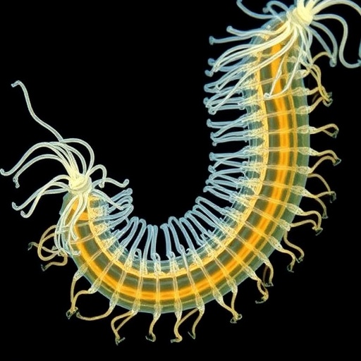
The extraordinary motility of Vibrio cholerae, the causative agent of cholera, is a key determinant of its lifecycle complexity and infectious potential. Central to this motility is a uniquely sheathed polar flagellum that rotates to propel the bacterium through aquatic and host environments. Although the structural composition of unsheathed flagella has long been explored, the enveloped and multi-component nature of the V. cholerae flagellum has posed significant challenges for high-resolution structural elucidation—until now. In an innovative study employing a synergetic combination of in situ cryo-electron microscopy (cryo-EM) single-particle analysis, fluorescence microscopy, and meticulously designed molecular genetics, researchers have unveiled the near-atomic level architecture of the sheathed flagellar filament, reshaping our understanding of its assembly and rotational mechanics.
At the core of this research lies the determination of remarkable structural resolutions ranging between 2.92 and 3.43 angstroms directly from intact V. cholerae cells, providing unprecedented insight into the spatial arrangement and interplay of the four integral flagellin proteins, FlaA through FlaD. These proteins do not simply serve redundant roles; instead, they orchestrate a highly ordered, cooperative assembly culminating in a filament that is structurally and functionally distinct from previously characterized unsheathed flagella. Notably, the study identifies FlaA as the pivotal scaffolding protein localized precisely at the bacterial cell pole, underpinning the nucleation and templating for the entire flagellar filament’s elaborate assembly process.
The flagellar filament’s sheath emerges as a truly unique feature of V. cholerae, presenting a membranous envelope continuous with the bacterium’s outer membrane. This membranous sheath encases the filament in a way rarely observed in bacterial motility structures, imparting physical and biochemical properties that are essential for the pathogen’s distinct modes of movement and environmental interaction. One of the most compelling discoveries from the researchers’ structural data is a highly conserved core filament architecture enveloped by a surprisingly smooth, hydrophilic surface. This surface likely facilitates intimate interactions with the sheath, reducing friction and mechanical resistance during filament rotation.
In contrast to unsheathed counterparts, the sheathed V. cholerae filament is characterized by an intricate surface chemistry tuned for a stable but dynamic interface with the sheath. The research posits that such adaptation is critical in enabling the filament to rotate as a free-standing entity within the membrane sheath, decoupling its motion from that of the sheath itself. This decoupling likely represents a significant evolutionary advantage, as it allows flagellum-driven propulsion without compromising integrity or imposing stress on the surrounding membrane.
The molecular basis for the filament’s supercoiling—a hallmark of directional motility and propulsion efficiency—was elegantly explained through subtle single-flagellin conformational changes uncovered in the high-resolution maps. These nanoscale rearrangements collectively translate into macroscopic supercoiling of the filament, inducing curvature in the surrounding membranous sheath. This supercoiled geometry not only optimizes hydrodynamics during bacterial swimming but also aligns with established theoretical models of flagellar propulsion in sheathed systems.
The use of in situ cryo-EM enabled visualization of the flagellar filament under near-native physiological conditions, circumventing artifacts associated with traditional sample preparation methods. This approach was essential for resolving the native arrangement of FlaA through FlaD subunits within the intact sheath environment, providing credence to the filament’s supramolecular assembly model. Complementary genetic manipulation confirmed the functional roles of the individual flagellins, validating the structural observations with phenotypic motility assays and fluorescence localization studies.
Further, the findings elucidate the dynamic interplay between the filament and sheath during rotation. Unlike models where the filament and sheath rotate in unison, the data suggest a sliding motion, where filament rotation generates propulsion while the sheath remains predominantly static, serving as a protective and stabilizing layer. This novel mechanism redefines paradigms of bacterial locomotion and points toward a sophisticated molecular machinery evolved for environmental resilience and host colonization.
Implications of this work extend beyond fundamental microbiology. Understanding the detailed architecture and mechanics of the V. cholerae flagellum provides critical targets for disruption of motility—a promising avenue for intervention aiming to attenuate pathogen virulence. Therapeutic strategies could be designed to destabilize sheath-filament interactions or inhibit flagellin assembly, potentially crippling the bacterium’s ability to reach and colonize host intestinal tissues.
Moreover, the structural principles unveiled could inspire biomimetic engineering applications. The unique membrane-sheathed, supercoiled filament capable of independent rotation suggests design blueprints for nanoscale rotary devices operating within confined lipid environments. Such bioinspired constructs could revolutionize targeted drug delivery systems or microscale swimmers for environmental remediation.
This comprehensive structural characterization also prompts reconsideration of how bacterial appendages evolve under selective pressures imposed by distinct niches. The presence of multiple flagellin types combined into a single filament may represent an evolutionary strategy to balance flexibility, robustness, and immune evasion. Investigations into homologous sheathed flagellar systems in other marine and pathogenic bacteria could reveal whether this architecture is a widespread adaptation or a specialized feature of Vibrio species.
Overall, this study stands as a testament to the power of integrating cryo-EM with genetic and biochemical tools to untangle complex bacterial nanomachinery. The resolution attained is pushing the boundaries of what can be resolved within living microbial cells, signaling a new era in structural microbiology. The insights gained not only deepen our molecular understanding of bacterial motility but also spotlight the intricate strategies microbes employ to thrive in diverse environments.
Future work will likely delve into the dynamic aspects of sheath and filament interactions during varying environmental stimuli, such as changes in osmotic pressure or host immune responses. Time-resolved cryo-EM and advanced fluorescence resonance energy transfer (FRET) studies may shed light on conformational plasticity and mechanical coupling underlying flagellar function. Additionally, exploring the regulatory networks controlling the expression and modification of FlaA-D proteins could reveal layers of control fine-tuning motility in response to environmental cues.
In conclusion, the structural revelations of the V. cholerae sheathed flagellum elucidate a finely tuned molecular device, expertly crafted through evolution to support bacterial locomotion and virulence. Its combination of a conserved core filament, multiple flagellin subunits, and a unique hydrophilic membranous sheath encasing the rotating filament embodies an elegant solution to the challenges of motile life in complex habitats. As such, this landmark work will undoubtedly inspire a wave of research focused on microbial motility, pathogenesis, and applied nanobiotechnology.
Subject of Research: The structural and functional mechanisms underpinning the assembly and rotation of the sheathed flagellar filament in Vibrio cholerae.
Article Title: Structures of the sheathed flagellum reveal mechanisms of assembly and rotation in Vibrio cholerae.
Article References:
Guo, W., Zhang, S., Park, J.H. et al. Structures of the sheathed flagellum reveal mechanisms of assembly and rotation in Vibrio cholerae. Nat Microbiol (2025). https://doi.org/10.1038/s41564-025-02161-x
Image Credits: AI Generated
Tags: advanced microscopy methodsaquatic bacterial movementcholera pathogen lifecyclecryo-electron microscopy techniquesflagellar assembly mechanismsflagellin protein interactionsinfectious disease researchmolecular genetics in microbiologyprotein structural resolutionsheathed flagellum structurestructural biology of bacteriaVibrio cholerae motility


