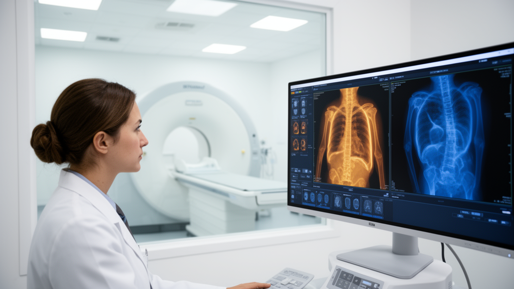
A shorter positron emission tomography scan could deliver the same diagnostic information as the lengthy protocols currently used for chest imaging, according to a retrospective analysis published in European Radiology Experimental. Researchers reconstructed PET data from 101 patients who underwent fluorine-18 fluorodeoxyglucose (18F-FDG) PET/MRI of the chest on a 3-Tesla integrated scanner and found that a 7-minute acquisition produced subjective image quality and lesion detectability comparable to a full 20-minute reference scan. For purely quantitative measurements, even shorter acquisitions of 3 to 5 minutes proved sufficient, and selected patients might be adequately assessed with as little as 2 minutes.
The study, conducted at a single center, took advantage of the way modern PET scanners store data. Rather than acquiring new scans, the team worked from list-mode files, the raw detection records of every positron event captured during the original 20-minute chest acquisition. From these files they retrospectively reconstructed ten simulated acquisition durations, ranging from 0.5 to 20 minutes, using identical reconstruction settings including time-of-flight information, point spread function correction, and ordered subset expectation maximization with 2 iterations and 28 subsets. This approach allowed a systematic, like-for-like comparison of what a radiologist would actually see had the scanner stopped earlier.
The patient cohort consisted of 77 men and 24 women with an average age of 63.8 years, drawn from an initial pool of 126 patients scanned between March 2022 and April 2023 after exclusions for MRI contraindications, absent fluorodeoxyglucose-avid chest lesions, recent antitumor therapy, substantial artifacts, or incomplete datasets. Most patients had lung cancer, with esophageal cancer as the second most common primary malignancy, and 89 of the 101 patients had pathological confirmation of their disease. Across the cohort, 283 lesions were identified on the 20-minute reference images, with between one and six lesions per patient.
Two readers with 16 and 14 years of experience independently rated subjective image quality on a five-point Likert scale, blinded to acquisition parameters and clinical information, with disagreements resolved by consensus. Consensus scores climbed from a median of 2, indicating limited diagnostic value, at 0.5 minutes to a median of 5, indicating excellent lesion discrimination without image noise, at 20 minutes. The improvement was significant up to 7 minutes, but from 7 minutes onward the scores plateaued, with no statistically significant differences among any of the durations between 7 and 20 minutes. Interobserver agreement improved in parallel, rising from substantial kappa values at short acquisitions to almost perfect agreement at 15 to 20 minutes; at 7 minutes the weighted kappa was 0.751.
The quantitative results told a more nuanced story. Lesion maximum and mean standardized uptake values, along with the lesion-to-background ratio, all decreased inversely with scan duration, a pattern the authors attribute to noise-induced bias. Very short acquisitions contain statistical noise that artificially inflates the maximum uptake value, so as acquisition time lengthens and noise falls, the measured values settle toward their true levels rather than deteriorating. Mean lesion uptake stabilized completely at 3 minutes, while the maximum uptake value and lesion-to-background ratio stabilized at 5 minutes. The image noise ratio of the background tissue required a full 10 minutes before further lengthening produced no significant differences. Metabolic tumor volume, by contrast, showed no significant variation across any acquisition time at all.
Lesion detectability provided perhaps the most clinically direct measure. The researchers defined detectability relative to the 20-minute reference, counting how many of the 283 reference-visible lesions were missed at each shorter duration. Miss rates rose steadily as scans shortened, reaching 7.77 percent, or 22 of 283 lesions, at 0.5 minutes, while a 15-minute acquisition missed none. At 7 minutes the miss rate was low, at 1.4 percent with a 95 percent confidence interval of 0.6 to 3.6 percent, and only 4 percent of patients had any missed lesion, with no patient missing two or more. At 5 minutes the miss rate nearly doubled to 2.5 percent and affected 7 percent of patients. Lesions missed at intermediate durations of 5 to 10 minutes were predominantly small nodules near the resolution limit of PET, where partial volume effects and limited count statistics are known constraints, while at very short acquisitions of 0.5 to 3 minutes high background noise was the dominant masking factor.
Subgroup analyses suggested that scan duration could be tailored to individual circumstances. When patients were stratified by the fluorodeoxyglucose uptake period before imaging, those with uptake of 80 minutes or less showed stable uptake parameters from 1.5 minutes onward, whereas patients with longer uptake periods of more than 80 minutes required 3 minutes. The authors offer a physiological explanation: a shorter uptake period may coincide with rapid tracer accumulation in tumors, when image contrast is already high, while longer uptake periods may involve the beginning of tracer clearance, yielding diminishing returns. Lesion size mattered as well. For lesions of 15 millimeters or larger, uptake parameters stabilized from 2 minutes onward, but lesions smaller than 15 millimeters, which are more vulnerable to partial volume effects and noise, required 3 minutes.
The findings carry practical weight for imaging departments. PET acquisition time in integrated PET/MRI is often set to match the duration of the simultaneous MRI sequences, which in this protocol meant 20 minutes of PET to align with roughly 20 minutes of chest MRI. Long scans test patient tolerance, and discomfort during extended acquisitions promotes motion, which itself degrades image quality. A 7-minute PET window would reduce motion artifacts, improve patient comfort, and increase scanner throughput, allowing more examinations per unit of tracer and machine time. The authors note that the 7-minute threshold represents the earliest point at which subjective quality matched the full protocol, and that the low miss rate and well-characterized causes of missed lesions at that duration support its use for routine diagnostic imaging, while quantitative tasks could be performed with 3 to 5 minutes.
Several limitations frame the conclusions. The study was retrospective, single-center, and conducted on a single scanner platform from GE Healthcare, so the specific thresholds may not transfer directly to systems from other manufacturers, although the underlying physical principles suggest similar trends should apply. The subgroup cutoffs, an 80-minute uptake period and lesion size categories, were data-driven, and the 20-minute reference served as the internal standard rather than an external gold standard such as pathological verification of every lesion. The small number of missed lesions precluded robust correlation analyses, and only standard tracer activity, averaging 4.11 megabecquerels per kilogram of body weight with a mean uptake period of about 66 minutes, was tested. The authors call for prospective, multicenter validation before the shortened protocols enter routine practice, but the central message stands: for chest 18F-FDG PET/MRI, most of the diagnostic value of a 20-minute scan appears to be captured in a fraction of the time.
Subject of Research: Retrospective study determining the minimum PET acquisition time for thoracic 18F-FDG PET/MRI that preserves image quality and lesion detectability
Article Title: Optimization of PET acquisition time for thoracic lesions in 18F-FDG PET/MRI: a retrospective analysis
Article References: Gu, H., Jiang, Y., Cai, J., Yang, G., Li, J., Chen, S., Luo, S., & Zhang, L. (2026). Optimization of PET acquisition time for thoracic lesions in 18F-FDG PET/MRI: a retrospective analysis. European Radiology Experimental, 10(1), Article 151. https://doi.org/10.1186/s41747-026-00787-y
Image Credits: AI Generated
DOI: 10.1186/s41747-026-00787-y
Keywords: PET/MRI, 18F-FDG, thoracic imaging, acquisition time, image quality, lesion detectability, SUV, lung cancer, radiology, scanner throughput, motion artifacts, retrospective study
News Source: Ophelia Keating. (October 29, 2026). Seven Minutes May Be Enough: Faster Chest PET/MRI Scans Match 20-Minute Image Quality. Scienmag.