Researchers develop a low-cost and easy-to-use cryogenic electron microscope.
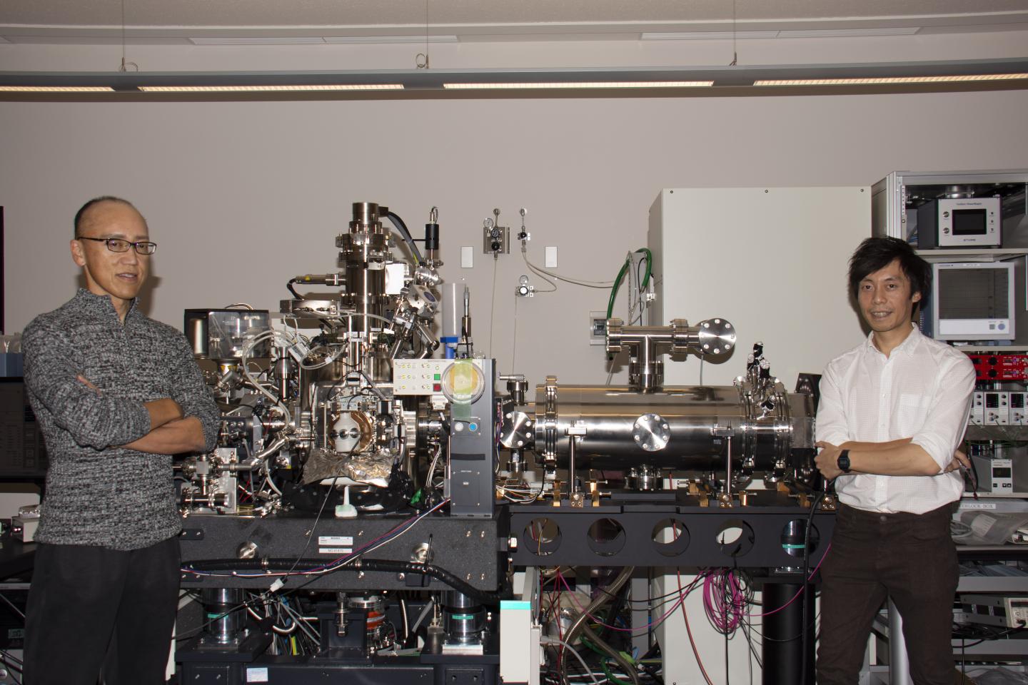
Credit: OIST
Visualizing the structure of viruses, proteins and other small biomolecules can help scientists gain deeper insights into how these molecules function, potentially leading to new treatments for disease. In recent years, a powerful technology called cryogenic electron microscopy (cryo-EM), where flash-frozen samples are embedded in glass-like ice and probed by an electron beam, has revolutionized biomolecule imaging. However, the microscopes that the technique relies upon are prohibitively expensive and complicated to use, making them inaccessible to many researchers.
Now, scientists from the Okinawa Institute of Science and Technology Graduate University (OIST) have developed a cheaper and more user-friendly cryo-electron microscope, which could ultimately put cryo-EM in reach of thousands of labs.
In a six-year construction process, the team built the microscope by adding a new imaging function to a scanning electron microscope. They used the hybrid microscope to image three different biomolecules: two distinctly shaped viruses and an earthworm protein.
“Building this microscope was a long and challenging process, so we are thrilled about its results so far,” said Dr. Hidehito Adaniya, a researcher in the Quantum Wave Microscopy (QWM) Unit and co-first author of the study, published in Ultramicroscopy. “As well as being cheaper and simpler to use, our microscope utilizes low-energy electrons, which could potentially improve the contrast of the images.”
Currently, cryo-EM works by firing high-energy electrons at a biological specimen. The electrons interact with atoms in the biomolecule and scatter, changing their direction. The scattered electrons then hit detectors, and the specific scatter pattern is used to build up an image of the sample.
But at high energies, only a relatively small number of these scattering events occur because the electrons interact very weakly with the atoms in the sample as they speed past. “Biomolecules are predominantly composed of elements with a low atomic mass, such as carbon, nitrogen, hydrogen and oxygen,” explained co-author and researcher, Dr. Martin Cheung. “These lighter elements are practically invisible to high-speed electrons.”
In contrast, low-energy electrons travel slower and interact more strongly with the lighter elements, creating more frequent scattering events.
This strong interaction between low-energy electrons and lighter elements is challenging to harness, however, because the layer of ice surrounding the specimen also scatters electrons, creating background noise that masks the biomolecules. To overcome this issue, the scientists adapted the microscope so it could switch to a different imaging technique: cryo-electron holography.
Forming the hologram
In holographic mode, an electron gun fires a beam of low-energy electrons towards the specimen so that part of the electron beam passes through the ice and specimen, forming an object wave, while the other part of the electron beam only passes through the ice, forming a reference wave. The two parts of the electron beam then interact with each other, like colliding ripples in a pond, creating a distinct pattern of interference – the hologram.
Based on the hologram’s interference pattern, the detectors can distinguish scattering by the specimen from scattering by the ice film. Scientists can also compare the two parts of the beam to gain extra information from the electrons that is difficult to detect using conventional cryo-EM.
“Electron holography provides us with two different kinds of information – amplitude and phase – whereas conventional cryo-electron microscopy techniques can only detect phase,” said Dr. Adaniya. This added information could allow scientists to gain more knowledge about the structure of the specimen, he explained.
A breakthrough in thin ice
In addition to building the hybrid microscope, the scientists also had to optimize the sample preparation. Since low-energy electrons are more prone to being scattered by the ice than high-energy electrons, the ice film enveloping the sample had to be as thin as possible to maximize the signal. The scientists used flakes of hydrated graphene oxide to hold the biomolecules in place, allowing thinner films of ice to form.
The scientists also had to take special steps to prevent the formation of crystalline ice, which is “bad news for cryo-EM imaging”, Cheung said.
With the current set up and optimized samples, the microscope produced images with a resolution of up to a few nanometers, which the researchers acknowledge is far lower than the near-atomic resolution achieved by conventional cryo-EM.
But even with the current resolution, the microscope still fills an important niche as a pre-screening microscope. “Because the low-energy electrons interact so strongly with the ice, our cheaper and user-friendly microscope can help researchers gauge their ice quality before spending valuable time and money using conventional cryo-EM microscopes,” said Dr. Adaniya.
The whole process is quick and simple, the researchers say. The SEM/STEM mode helps scientists locate the best spot for imaging, followed by a seamless transition into the holographic mode. What’s more, the ability for this mode-switching technology to be implemented into other commercial scanning electron microscopes makes it a widely adoptable imaging method.
In the future, the team hopes to improve image resolution further, by changing the electron gun to one that creates a higher quality electron beam. “That will be the next step forward,” they said.
###
Media Contact
Tomomi Okubo
[email protected]
Original Source
https:/
Related Journal Article
http://dx.

