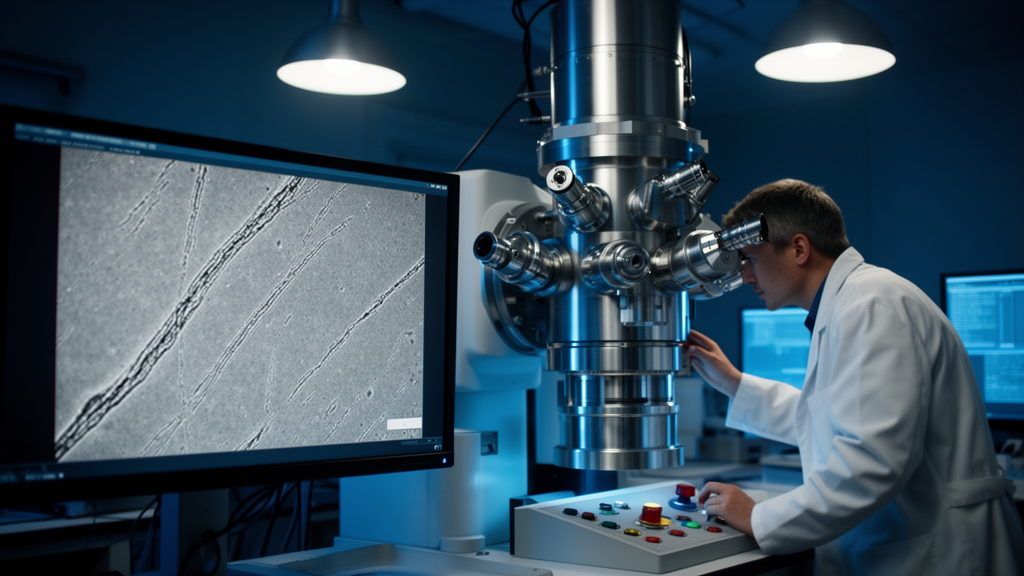
Electron microscopy can reveal the finest architecture of cells and tissues, but the images it produces are rarely pristine. Biological samples must be fixed, stained, dehydrated and embedded so they can survive the high vacuum of the microscope chamber and the relentless bombardment of the electron beam, and every step of that preparation can leave traces that masquerade as biology. A team at Delft University of Technology and the University Medical Center Groningen has now catalogued, artifact by artifact, what goes wrong when a relatively new detection technique called optical scanning transmission electron microscopy, or OSTEM, is combined with conventional sample preparation protocols, and they show that with careful controls each artifact can be suppressed or eliminated.
OSTEM is designed for a specific challenge: imaging very large areas, and eventually volumes, of biological tissue at nanometer resolution. In conventional scanning electron microscopy, images are built from electrons that bounce off or are emitted from the sample surface. In transmission modes, electrons that pass through an ultrathin section are collected instead. OSTEM takes an unusual route: the section is deposited directly onto a scintillator, a crystal that converts transmitted electrons into flashes of light. A high numerical aperture objective beneath the crystal collects those photons and projects them onto a multipixel photon counter. Because the technique works with both single-beam instruments and multibeam platforms such as the commercial FAST-EM, which speeds up acquisition by scanning many beamlets in parallel, it promises large-scale two-dimensional and three-dimensional datasets that would be impractically slow to collect otherwise.
The price of that design is that the electron beam does not stop at the tissue. Its interaction volume extends through a thin metallic coating layer and into the scintillator itself, so every imperfection between the substrate and the sample is superimposed directly on the image of the tissue. The researchers found that this makes OSTEM unusually revealing of flaws that conventional detection schemes would never register, and in some cases introduces artifacts of its own.
The first artifact concerns staining chemistry. When ultrathin sections of zebrafish larva tissue were post-stained with neodymium acetate after being mounted on the scintillator, OSTEM images showed darker patches that were absent from images taken with backscattered electron detection at low landing energy. When the landing energy was raised to match that used in OSTEM, the same patches appeared even in backscattered images. The artifacts were not seen outside the section, even though staining solution had been applied there too. Together, these observations pointed to a specific origin: residual staining solution trapped beneath the section at the interface with the substrate, drawn under the tissue by capillary force. En bloc staining, in which the stain is applied before sectioning, avoided the problem entirely.
A second family of artifacts arose from the coating itself. Scintillators used in the study carry a nanometer-scale molybdenum film that acts as a spacer, conducts away charge and heat, and reflects emitted photons back toward the objective. When rat pancreas tissue prepared with a reduced osmium-thiocarbohydrazide-osmium staining protocol was deposited on molybdenum-coated scintillators using a loop transfer through a water droplet, fractal-like electron-dense precipitates formed on and under the sections. These structures resembled the branching patterns produced by diffusion-limited aggregation, suggesting a reaction between the molybdenum coating and a compound in the staining protocol, mediated by the aqueous medium. Several observations supported this interpretation: the precipitates were not continuous across serial sections, so they must have formed after embedding, most likely during section collection; they did not form on coatings of other materials such as chromium or graphene; and they appeared mostly over tissue rather than empty resin. When the scintillator was instead submerged in the water bath of the diamond knife and dried by lowering the water level, the precipitates did not form, apparently because the large volume of bath water diluted any reaction product.
Coating quality itself proved decisive. High-quality sputtered molybdenum films have a homogeneous grey reflective surface, but some scintillators developed drying rings or became more transparent after exposure to water or air, changes consistent with oxidation of the molybdenum, whose oxide reflects far less light than the metal. On a scintillator with a visibly degraded film, the contrast of the biological sample in large-scale acquisitions was clearly affected, although fine structure remained discernible. The authors recommend visual inspection of every substrate both before and after specimen preparation so that compromised coatings are caught before they ruin an experiment.
Perhaps the most quantifiable artifact came from the substrate surface itself. Because the beam probes the scintillator, scratches, pits and polishing marks on its surface all contribute to the image and dilute the contrast of the biology. Atomic force microscopy of molybdenum-coated yttrium aluminum garnet scintillators revealed large roughness differences between batches and suppliers. Two scintillators carrying tissue samples differed roughly twofold in root-mean-square roughness, about 5 nanometers versus about 2.5 nanometers, and the biological image from the rougher substrate was visibly grainier: nuclear membranes and mitochondrial cristae that could be resolved on the smoother substrate were lost on the rougher one. Control experiments with backscattered detection at increasing landing energies showed that image quality diverged between the two samples only once the interaction volume extended into the substrate, confirming that surface irregularities, not preparation differences, were responsible. The best substrates, from a supplier called Surface Preparation Laboratory and from an optimized batch, showed roughness below 1 nanometer and no scratch marks, and scintillators of that quality have delivered high-quality biological datasets.
Even the manufacturing history of the crystal leaves fingerprints. Without additional broad-ion-beam polishing after crystal growth, scintillator surfaces carry micrometer- and nanometer-scale scratches that show up in OSTEM images. Polishing removes those scratches but leaves dimple-shaped structures several micrometers across, which appear in multibeam datasets through the tissue as gentle elevations that obscure ultrastructure at their edges without blocking the transmission signal. The crystal growth process itself can also produce quasi-periodic intensity ripples, known as growth striations, arising from variations in dopant ion concentration; these appeared only on polished sides of the crystals and were absent in scintillators from the higher-quality supplier. Fortunately, the authors note, both dimples and striations are typically rare and homogeneous enough that interpretation of the ultrastructure is negligibly affected.
Multibeam operation adds artifacts of its own. Because adjacent beamlets scan overlapping regions, some areas of the sample receive double or quadruple doses, producing intensity differences in overlapping zones that are most likely explained by shrinkage or mass loss of the resin under irradiation. When the same region was scanned repeatedly, those intensity differences diminished over the first few scans, but from the fifth scan onward beam damage emerged as lines along the scan directions, most pronounced at corners exposed four times per scan, along with darkened edges attributed to carbon deposition. A separate quirk of the optical system, signal spillover between neighboring beamlets, creates phantom images: a high-contrast feature such as a dirt particle or a section fold attenuates the transmission signal and thereby alters the baseline spillover reaching adjacent detectors, conjuring a faint ghost of the feature in the neighboring field. Image post-processing can make matters worse. The standard correction, which averages all field images and subtracts the average from each, incorporates artifact-corrupted fields into that average and can imprint a background texture across the corrected data; excluding corrupted fields or using a median image avoids the problem.
The payoff of this systematic diagnosis is a practical one. By combining en bloc staining, substrate submersion during sectioning, quality-controlled scintillators with low roughness and stable coatings, optimized optical alignment to minimize spillover, and careful post-processing, the team acquired a large FAST-EM dataset of rat pancreas in which none of the identified artifacts were apparent, with mitochondrial cristae, insulin granules and collagen in the extracellular matrix clearly resolved. At that point, they conclude, the contrast of small biological features is limited by the sensitivity of OSTEM detection itself rather than by substrate flaws. For a technique aimed at mapping whole sections and volumes of tissue, knowing exactly where the ghosts come from, and how to banish them, is what turns an intriguing detection scheme into a reliable tool for unbiased, large-scale imaging of biological structure.
Subject of Research: Artifacts in optical scanning transmission electron microscopy of biological samples and their mitigation
Article Title: Mitigation of artifacts in imaging biosamples with optical scanning transmission electron microscopy
Article References: Kievits, A. J., Duinkerken, B. H. P., Lane, R., Giepmans, B. N. G., & Hoogenboom, J. P. (2026). Mitigation of artifacts in imaging biosamples with optical scanning transmission electron microscopy. Applied Microscopy, 56(1), Article 11. https://doi.org/10.1186/s42649-026-00132-y
Image Credits: AI Generated
DOI: 10.1186/s42649-026-00132-y
Keywords: electron microscopy, OSTEM, artifacts, scintillator, sample preparation, multibeam imaging, FAST-EM, molybdenum coating, surface roughness, beam damage, signal spillover, volume electron microscopy
News Source: Denise Maddox. (October 29, 2026). Seeing the Flaws: New Map of Artifacts in Optical Scanning Electron Microscopy. Scienmag.