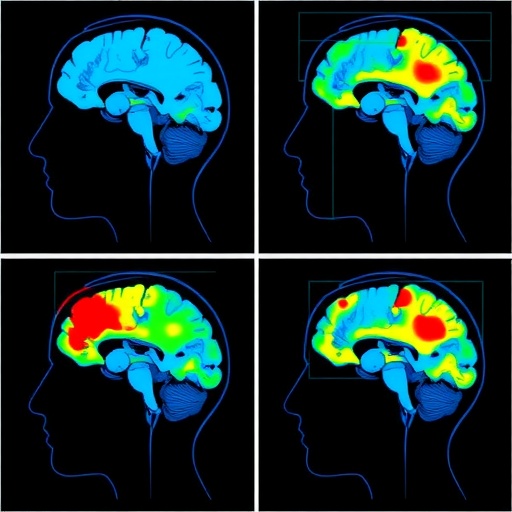
Swallowing is one of those bodily functions most people perform hundreds of times a day without a second thought, yet it depends on a remarkably intricate dialogue between the brainstem, the motor cortex and a distributed web of higher-order cortical regions. When that dialogue breaks down—as it often does after stroke, traumatic brain injury or in neurodegenerative disease—the result is dysphagia, a swallowing impairment that can lead to choking, aspiration pneumonia, malnutrition and a dramatic decline in quality of life. Now, a comprehensive scoping review published in Biomedical Engineering Letters has mapped two decades of research using a portable, light-based brain imaging technology called functional near-infrared spectroscopy, or fNIRS, to watch the human cortex at work during swallowing. The review, conducted by Na-Kyoung Hwang of Seoul Metropolitan Bukbu Hospital, Gihyoun Lee of Chonnam National University and Ji-Su Park of Pusan National University, offers both an encouraging verdict and a sobering caveat: the technology works, but the field has yet to agree on how to use it.
The appeal of fNIRS lies in its simplicity and its tolerance of real-world behavior. Unlike functional magnetic resonance imaging, which requires participants to lie motionless inside a loud, cramped scanner, fNIRS relies on nothing more intimidating than a cap fitted with light emitters and detectors pressed gently against the scalp. Near-infrared light at wavelengths around 700 to 900 nanometers penetrates the skull and skull-adjacent tissues by a few centimeters, and because oxygenated and deoxygenated hemoglobin absorb those wavelengths differently, the device can calculate changes in cortical blood oxygenation from the fraction of light that scatters back to the detectors. When a brain region becomes more active, local blood flow increases and delivers a surplus of oxygenated hemoglobin—a signal fNIRS can track with a temporal resolution of fractions of a second. That makes the technique well suited to swallowing, a behavior that unfolds in under two seconds and that cannot be cleanly performed inside an MRI bore, particularly by frail or elderly patients.
To chart how researchers have exploited this window onto the swallowing brain, the Korean team followed the Joanna Briggs Institute methodology for scoping reviews and adhered to the PRISMA-ScR reporting guidelines. They searched four major databases—MEDLINE, Embase, Scopus and Web of Science—from their inception through September 2025, casting a wide net across experimental and clinical literatures. After screening, 21 studies met the inclusion criteria: investigations involving adult participants in whom fNIRS was used to measure swallowing-related cortical activity, whether during natural swallowing tasks, deliberately designed swallowing exercises, sensory stimulation paradigms or neuromodulation interventions. The studies spanned healthy volunteers as well as clinical populations, most notably stroke survivors with dysphagia.
The central finding to emerge from the synthesis is strikingly consistent: swallowing recruits a distributed fronto-sensorimotor cortical network rather than a single “swallowing center.” Across studies, hemodynamic responses clustered over the primary motor and sensory cortices—particularly the regions representing the tongue, pharynx and larynx—along with the inferior frontal gyrus, the supplementary motor area and adjacent prefrontal territory. This pattern aligns with the classical model of swallowing control, in which the brainstem central pattern generator executes the reflexive sequence of muscle contractions while cortical areas handle the volitional initiation, sensory gating and fine tuning of the swallow. The reviews’ authors note that the degree and spatial distribution of this activation are not fixed: they shift depending on what the swallow entails, what sensations accompany it and what the brain is being asked to do differently.
One of the most clinically provocative threads in the reviewed literature concerns sensory modulation. Several studies examined how manipulating the oral environment changes cortical responses to swallowing. Acidic solutions, for example, produced stronger and sometimes more bilateral cortical activation than plain water, suggesting that sour taste stimuli heighten the salience of the swallow and amplify sensory feedback to the cortex. Other experiments delivered tactile vibration over the larynx or applied pharyngeal electrical stimulation and observed increases in both swallowing frequency and activation of the sensorimotor cortex. In patients with brainstem stroke—a group whose dysphagia is traditionally attributed to damage of the reflex machinery itself—visual and gustatory stimuli were still able to reshape cortical hemodynamic patterns, hinting that even “brainstem” dysphagia retains a cortical dimension that might be therapeutically targeted. For rehabilitation specialists, these findings support a long-standing hypothesis that intensifying sensory input during swallowing can drive use-dependent plasticity in the cortical networks that support recovery.
Motor demand tells a complementary story. Studies that compared simple water swallows with effortful or resistance-based swallowing exercises—such as the chin-tuck-against-resistance maneuver, in which patients swallow while flexing the neck against opposing force—found distinct cortical signatures associated with increasing motor challenge. This resonates with the “challenge point” framework from motor learning research, which holds that practice conditions must be sufficiently demanding to induce adaptation without overwhelming the learner. In practical terms, the reviewed evidence suggests that fNIRS could serve as a physiological dosimeter, allowing clinicians to verify that a given swallowing exercise is actually engaging the intended cortical circuitry in a given patient, rather than relying on surface muscle activity alone.
Perhaps the most forward-looking section of the review concerns neuromodulation. Several included studies paired fNIRS with non-invasive brain stimulation techniques. Repetitive transcranial magnetic stimulation, delivered over the swallowing motor cortex, was shown in randomized studies of stroke patients to alter hemodynamic signals in parallel with measurable swallowing improvements. Transcranial direct current stimulation experiments compared stimulation amplitudes over the sensorimotor cortex and documented dose-dependent cortical responses. Modified pharyngeal electrical stimulation changed swallowing-related functional connectivity, and even electroacupuncture at traditional acupuncture points produced stronger activation of the swallowing cortex than single-point stimulation. Together, these studies sketch a future in which fNIRS is not merely an observational tool but a real-time feedback instrument—confirming within a single session whether a neuromodulatory intervention is shifting cortical excitability in the desired direction, and potentially enabling closed-loop rehabilitation protocols in which stimulation parameters are titrated against the patient’s own cortical response.
Yet the review is equally emphatic about what stands in the way of that future. Methodological heterogeneity pervades nearly every aspect of the field. Studies differ in task paradigms—dry swallows versus water swallows versus flavored liquids; single swallows versus repeated blocks; volitional versus spontaneous swallows—each of which is known to elicit somewhat different hemodynamic signatures. Optode placement varies widely, with some groups densely sampling the sensorimotor strip and others covering broader prefrontal territories, which complicates any direct comparison of activation maps. Analytical pipelines are similarly divergent, encompassing different filtering choices, baseline corrections, channel-averaging schemes and statistical thresholds. The reviewers also flag a more fundamental imbalance: of the 21 studies, the majority enrolled healthy adults, and the number of investigations involving actual dysphagia patients—particularly longitudinal studies tracking recovery—remains small. Without larger clinical cohorts and standardized measurement protocols, the field cannot yet determine whether cortical activation patterns measured by fNIRS predict which patients will recover swallowing function or respond to particular therapies.
The implications for rehabilitation are nonetheless considerable. Dysphagia affects a substantial proportion of stroke survivors and is a leading contributor to post-stroke aspiration pneumonia, yet bedside and instrumental assessments—videofluoroscopy and fiberoptic endoscopic evaluation—capture the mechanical act of swallowing rather than the neural drive behind it. fNIRS, by contrast, is inexpensive, portable, silent and compatible with bedside use, making it conceivable that future dysphagia assessments could incorporate a cortical activation profile alongside structural and functional measures. Such profiles could reveal, for example, whether a patient’s swallowing network has shifted activation toward the undamaged hemisphere after a stroke—a pattern of reorganization that neuroimaging research in limb motor recovery has linked to better outcomes. The reviewers point to earlier fMRI work demonstrating time-dependent hemispheric shifts in swallowing control as a conceptual template that fNIRS could now pursue at the bedside and at much lower cost.
The path from scoping review to clinical standard is, of course, long. The authors call for the development of standardized fNIRS protocols for swallowing research—harmonized optode montages, task paradigms, data processing pipelines and clinically relevant outcome measures—so that findings from different laboratories and hospitals can be pooled and translated into practice. They also emphasize that fNIRS has inherent limitations: light penetrates only the outer cortex, leaving brainstem and subcortical swallowing centers invisible to the technique, and signals can be contaminated by scalp blood flow, head motion and the very jaw and neck movements that swallowing entails. Sophisticated signal processing and careful experimental design will be needed to disentangle genuine cortical hemodynamics from these artifacts.
Still, the overall message of the review is one of cautious optimism. Twenty-one studies have collectively demonstrated that fNIRS is feasible and sensitive enough to detect swallowing-related cortical activation in both experimental and clinical settings, and that the measured signals respond meaningfully to sensory stimulation, motor demands and neuromodulation. What the field lacks is not proof of principle but coordination. If researchers can converge on shared protocols and expand their focus to the clinical populations who stand to benefit most, the humble near-infrared cap—already a fixture in stroke rehabilitation research and brain-computer interface laboratories—could become a routine instrument in the dysphagia clinic, transforming swallowing rehabilitation from an empirical craft into a neuroscientifically guided therapy. For the millions of patients who struggle to swallow safely each day, bringing the swallowing brain into view may be the first step toward rewiring it.
Subject of Research: Cortical activation during swallowing-related tasks measured with functional near-infrared spectroscopy (fNIRS), and its implications for dysphagia assessment and rehabilitation.
Subject of Research: Technology and Engineering
Article Title: fNIRS-based cortical activation during swallowing-related tasks: implications for dysphagia rehabilitation—a scoping review
Article References: Hwang, N.-K., Lee, G., & Park, J.-S. (2026). fNIRS-based cortical activation during swallowing-related tasks: implications for dysphagia rehabilitation—a scoping review. Biomedical Engineering Letters. https://doi.org/10.1007/s13534-026-00601-z
Image Credits: AI Generated
DOI: 10.1007/s13534-026-00601-z
Keywords: Dysphagia, Swallowing, Functional near-infrared spectroscopy, Cortical activation, Neurorehabilitation, Neuromodulation, Stroke, Sensorimotor cortex, Hemodynamic response, Scoping review
Cite Scienmag News
APA
MLA
Chicago
Cassandra Pierce. (September 9, 2026). Scoping review maps cortical activation during swallowing tasks using fNIRS. Scienmag. https://scienmag.com/scoping-review-maps-cortical-activation-during-swallowing-tasks-using-fnirs/
Cassandra Pierce. “Scoping review maps cortical activation during swallowing tasks using fNIRS.” Scienmag, 9 September 2026, https://scienmag.com/scoping-review-maps-cortical-activation-during-swallowing-tasks-using-fnirs/. Accessed 9 September 2026.
Cassandra Pierce. “Scoping review maps cortical activation during swallowing tasks using fNIRS.” Scienmag. September 9, 2026. https://scienmag.com/scoping-review-maps-cortical-activation-during-swallowing-tasks-using-fnirs/
Copy citation
Download RIS
Tags: advances in functional near-infrared spectroscopybrain imaging for dysphagia assessmentbrain-muscle communication in swallowingbrainstem and cortical involvement in swallowingchallenges in fNIRS application for swallowing studieschallenges in neuroimaging standardizationcortical activation during swallowingfNIRS brain imaging in dysphagiafNIRS in neurorehabilitationfunctional near-infrared spectroscopy in neurorehabilitationmapping brain activity during swallowing tasksneural correlates of swallowingneurodegenerative disease and swallowing impairmentneuroimaging of swallowing disordersneuroplasticity in swallowing recoveryportable brain imaging technologiesportable neuroimaging technologies for swallowingreal-world applications of fNIRSreal-world brain imaging techniquesstroke-related dysphagia assessment