


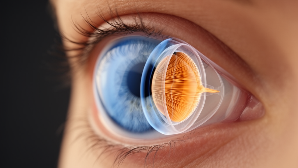
For the first time, researchers have applied digital image correlation (DIC) to optical coherence tomography images to measure, in living eyes, the internal strain that builds up inside the ciliary muscle when it contracts during accommodation. The study, published in Eye and Vision, describes a validated framework that goes beyond the traditional practice of measuring how the muscle thickens or moves forward, and instead extracts pixel-level deformation fields from the speckle patterns that OCT images naturally contain. In a proof-of-concept experiment involving 23 subjects, the method revealed a graded decline in effective strain across a control group, a high myopia group, and an elderly group, with the mean equivalent strain falling from 0.28 ± 0.06 in young controls to 0.20 ± 0.03 in high myopes and 0.16 ± 0.05 in older participants under a −5.0 diopter accommodative stimulus (overall P = 0.002).
The ciliary muscle is the engine of accommodation, the physiological process that reshapes the crystalline lens so the eye can focus on near objects. Its biomechanical properties are thought to be central to two of the most common visual conditions of modern life: myopia progression and presbyopia. Until now, however, OCT studies of the muscle have relied almost entirely on morphological surrogates, such as maximum ciliary muscle thickness and anterior ciliary muscle length, which convey anatomical change but say nothing about how the tissue deforms internally. Direct in vivo assessment of displacement and strain distribution across different regions of the muscle has remained largely out of reach.
DIC is a non-contact, full-field optical technique that tracks the displacement of random speckle patterns to calculate surface deformation with sub-pixel precision. Originally developed for micron-scale strain analysis of engineering materials, it has more recently found biomedical applications in bone deformation, vascular wall strain, and corneal biomechanics. The key insight of the new study is that OCT images inherently generate speckle patterns that can serve as natural texture signatures for DIC tracking, eliminating the need for artificial markers. The researchers used structural intensity images rather than phase-sensitive OCT, because commercial devices typically restrict access to raw phase data. Although intensity-based tracking is inherently less precise than phase-sensitive optical coherence elastography, it circumvents the need for custom-built hardware and makes the method broadly accessible on standard commercial OCT systems.
The study unfolded in three phases. In the first, a technical validation step, ciliary muscle images were acquired from two participants in a non-accommodative state using two swept-source OCT systems, the CASIA2 (Tomey, Japan, 1310 nm wavelength, 50,000 A-scans per second) and the VG200D (SVision Imaging, China, 1050 nm, 200,000 A-scans per second), each configured with four scanning modes based on image-averaging repetitions (R1, R8, R16, and R64). The segmented muscle regions were then subjected to artificial deformation simulations in MATLAB, with two deformation levels applied: a large condition of 200 µm horizontal compression and 100 µm vertical stretch, and a small condition of 40 µm horizontal compression and 10 µm vertical stretch. The open-source ncorr DIC algorithm processed the simulated images, and identification errors were calculated against the known inputs.
The CASIA2 system exhibited markedly lower error variability and superior detection stability compared with the VG200D, which the authors attribute partly to its 1310-nm wavelength offering better tissue penetration and contrast for speckle tracking. Among the scanning modes, R8 proved the most stable configuration. Un-averaged R1 images occasionally showed marginally lower errors for the control group but were highly susceptible to random shot noise in the high myopia group, where tissue scattering and signal-to-noise ratios are altered. Excessive averaging at R16 and R64 induced motion blur and over-smoothed the high-frequency speckle patterns needed for accurate cross-correlation. R8 therefore struck the optimal balance between noise suppression and speckle preservation, and it was adopted for all subsequent in vivo experiments.
The second phase tested the algorithm under realistic physiological noise using a cross-image strategy. Images were captured twice, 30 seconds apart, from three participants representing the control, high myopia, and elderly profiles. One image from each pair was digitally modified to carry the same deformation magnitudes used in the simulation phase, and the ncorr-DIC algorithm computed displacement and strain differences between the modified and unaltered images. As expected, tracking errors rose compared with the idealized simulations, providing direct evidence of natural speckle decorrelation between consecutive in vivo acquisitions. Nevertheless, the average detection error for small deformations was approximately 20 µm, confirming that the method retains favorable anti-noise capability and repeatability under realistic speckle conditions.
The third phase applied the validated protocol to 23 adults aged 20 to 50 years, divided into a control group of seven young adults with spherical equivalent refraction between −3.00 and +0.50 D, a high myopia group of seven with spherical equivalent ≤ −6.0 D or axial length ≥ 26.5 mm, and an elderly group of nine aged approximately 50 years. Accommodation was stimulated with a custom-built Badal optical system coupled to the OCT, in which translating the visual target forward by 5.625 mm produced a 1 diopter change in vergence. Images were acquired twice in the non-accommodative baseline state and once under a −5.0 D stimulus. After image registration anchored to the scleral spur, the modified ncorr implementation, which incorporated automated region-of-interest detection, yielded horizontal and vertical displacement vectors plus horizontal normal, shear, and vertical normal strain components, summarized by a von Mises-style equivalent strain.
The results showed that the method tracked deformation efficiently, with region recognition rates ranging from 81% to 98%, and that baseline displacement and strain under the two non-accommodative acquisitions were extremely close to zero with no significant differences among groups. Under the −5.0 D stimulus, the equivalent strain demonstrated the stepwise attenuation described above, driven primarily by horizontal compressive strain and vertical tensile strain, both diminished in the high myopia and elderly groups, while internal shear strain remained minimal and showed no statistically significant differences (P > 0.05). Morphological measurements corroborated the biomechanical findings: the control group showed pronounced anterior shortening (ΔCMAL = −102.43 ± 31.99 µm) and maximal thickening (ΔCMTMAX = 93.86 ± 29.91 µm) upon accommodation, while accommodative thickening was significantly reduced in the other two groups (overall Kruskal–Wallis, P = 0.003). A combined correlation analysis found the mean equivalent strain significantly positively correlated with macroscopic muscle thickening (r = 0.70, P < 0.001) and with accommodation amplitude (r = 0.69, P < 0.001).
The authors emphasize that this is a pilot study focused on methodological validation rather than definitive physiological conclusions. The small sample size limits generalizability, and larger multicenter cohorts will be needed to confirm population-level consistency and establish normative databases. The analysis used only a single −5.0 D stimulus, without dose-response or dynamic time-series analysis, and maintaining OCT speckle stability remains challenging, with noise differences between acquisitions causing some instability in displacement and strain results. The reliance on 2D cross-sectional B-scans to evaluate an inherently 3D sphincter-like contraction imposes theoretical limits, since out-of-plane motion induces speckle decorrelation, and anterior segment OCT carries geometric optical distortions that future ray-tracing corrections should address.
Even with those caveats, the framework marks a transition from static morphological observation to dynamic functional assessment of the eye’s focusing machinery. By quantifying mechanical attenuation associated with high myopia and age-related decline, DIC-based OCT analysis offers a new functional biomarker for investigating the mechanisms of accommodation dysfunction. The authors suggest it could prove valuable for the early detection of presbyopia, the objective assessment of anti-presbyopic interventions, and the quantitative exploration of accommodative function’s role in the onset and progression of myopia, provided future large-scale studies build on the quantitative foundation this proof-of-concept establishes.
Subject of Research: A DIC-based OCT method for measuring in vivo ciliary muscle deformation and strain during accommodation
Article Title: In vivo biomechanical property detection of the ciliary muscle based on optical coherence tomography and digital image correlation technology
Article References: Wang, J., Luo, S., Li, Y., Yi, S., Xu, N., Wang, T., Gao, Y., Yan, R., Lu, F., Wang, J., & Shao, Y. (2026). In vivo biomechanical property detection of the ciliary muscle based on optical coherence tomography and digital image correlation technology. Eye and Vision, 13(1), Article 28. https://doi.org/10.1186/s40662-026-00498-w
Image Credits: AI Generated
DOI: 10.1186/s40662-026-00498-w
Keywords: ciliary muscle, optical coherence tomography, digital image correlation, accommodation, myopia, presbyopia, strain, biomechanics, speckle tracking, swept-source OCT, ocular imaging, vivo
News Source: Ophelia Keating. (October 30, 2026). Scientists Track the Ciliary Muscle’s Hidden Strain During Accommodation. Scienmag.



