Roughly 100 trillion connections between neurons make it possible for the brain to function. Psychology Professor Brian Wandell’s group has devised a technique for mapping these connections with greater accuracy than ever before.
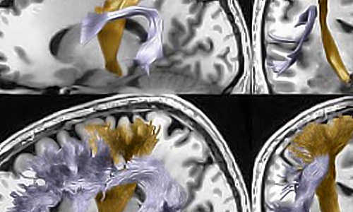
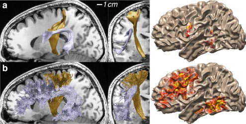
Two different algorithms produce two very different estimates of the shape of the same white matter connection in Franco Pestilli’s brain. The LiFE software, developed in the Wandell Lab, aims to produce more precise estimates. Photo Credit: Wandell Lab / Stanford
To see, think or feel, the 100 billion neurons in our brain must exchange messages. These are transmitted over some 100 trillion specialized connections, known collectively as the “connectome.” Most connections are extremely short, carrying information a few hundred-thousandths of an inch between nearby neurons. But many important connections are much longer, winding as much as a foot from one end of the brain to the other.
Scientists at Stanford University have developed a mathematical and computational technology that allows researchers to more accurately map the large, long connections within the white matter tissue of living human brains. The methodology is called LiFE, for linear fascicle evaluation. The work is detailed online in Nature Methods.
To test the new method, lead author Franco Pestilli analyzed MRI scans of two such connections within his own brain. In one analysis, the two structures – the arcuate fasciculus, which is involved in reading and language, and the corticospinal tract, which plays a role in motor coordination – appeared short and fairly smooth. In the other, the ridged tendrils within the tracts spread far longer and wider.
“Previously, scientists had no method for deciding which of the two representations of the human brain is correct,” said Pestilli, a research associate in the laboratory of Brian Wandell, a professor of psychology at Stanford. “As a result, different research groups using the same data often came to different conclusions. The new technology provides a mathematical analysis and open-source software to decide which of the two estimates is better.”
Many candidate representations of the human connectome can be created using available fiber tracking technology. LiFE interprets these pathways and uses them to simulate synthetic MRI signals. Candidate connectomes can be evaluated to find the one that generates a synthetic signal that most closely matches the actual data.
“I like to think about each candidate connectome as a prototype,” Pestilli said. “We can learn much by building many prototypes and finding the one that best represents the measured MRI signal. Once we have this optimized prototype, we can more accurately study the function of the actual pathways.”
The National Institutes of Health has made mapping the human brain an important scientific objective because of its importance in predicting healthy development and critical brain function. For example, in a series of papers over the years, the Wandell lab has shown that the properties of the brain connections differ between good and poor readers, particularly in one specific pathway, the arcuate fasciculus.
“We can much more reliably identify the arcuate fasciculus in a child who is struggling to read and understand whether or not that connection might be the source of the difficulty,” Wandell said.
Understanding the human connectome will allow clarifying the fundamental organizing principles of structure and function of the human brain. This will allow us to understand the mechanisms behind some of the most important diseases affecting individuals and society. For example, multiple sclerosis, Alzheimer’s disease and schizophrenia have all been regarded as brain diseases affecting the connectome.
An overall effort of the Wandell lab involves sharing data and computational methods with the broader scientific community. This work, which was funded by a grant from the National Science Foundation, supports that effort by providing data and a complete implementation of the method through the Stanford Digital Repository and GitHub.
“We hope other investigators use the code and improve it,” Pestilli said. “Code and data sharing can be a very important contribution as we try to understand the human brain.”
Story Source:
The above story is based on materials provided by Stanford University.


