• HOME
  • NEWS
  • EXPLORE
    • CAREER
      • Companies
      • Jobs
    • EVENTS
    • iGEM
      • News
      • Team
    • PHOTOS
    • VIDEO
    • WIKI
  • BLOG
  • COMMUNITY
    • FACEBOOK
    • INSTAGRAM
    • TWITTER
Sunday, February 1, 2026
BIOENGINEER.ORG
No Result
View All Result
  • Login
  • HOME
  • NEWS
  • EXPLORE
    • CAREER
      • Companies
      • Jobs
        • Lecturer
        • PhD Studentship
        • Postdoc
        • Research Assistant
    • EVENTS
    • iGEM
      • News
      • Team
    • PHOTOS
    • VIDEO
    • WIKI
  • BLOG
  • COMMUNITY
    • FACEBOOK
    • INSTAGRAM
    • TWITTER
No Result
View All Result
Bioengineer.org
No Result
View All Result
Home NEWS Science News Tissue Engineering

Scientists grow leg muscle from cells in a dish

Bioengineer by Bioengineer
February 25, 2015
in Tissue Engineering
Reading Time: 2 mins read
4
Share on FacebookShare on TwitterShare on LinkedinShare on RedditShare on Telegram

A team of researchers from Italy, Israel and the United Kingdom has succeeded in generating mature, functional skeletal muscles in mice using a new approach for tissue engineering. The scientists grew a leg muscle starting from engineered cells cultured in a dish to produce a graft. The subsequent graft was implanted close to a normal, contracting skeletal muscle where the new muscle was nurtured and grown. In time, the method could allow for patient-specific treatments for a large number of muscle disorders. The results are published in EMBO Molecular Medicine.

tissue

The scientists used muscle precursor cells – mesoangioblasts – grown in the presence of a hydrogel (support matrix) in a tissue culture dish. The cells were also genetically modified to produce a growth factor that stimulates blood vessel and nerve growth from the host. Cells engineered in this way express a protein growth factor that attracts other essential cells that give rise to the blood vessels and nerves of the host, contributing to the survival and maturation of newly formed muscle fibres. After the graft was implanted onto the surface of the skeletal muscle underneath the skin of the mouse, mature muscle fibres formed a complete and functional muscle within several weeks. Replacing a damaged muscle with the graft also resulted in a functional artificial muscle very similar to a normal Tibialis anterior.

Tissue engineering of skeletal muscle is a significant challenge but has considerable potential for the treatment of the various types of irreversible damage to muscle that occur in diseases like Duchenne muscular dystrophy. So far, attempts to re-create a functional muscle either outside or directly inside the body have been unsuccessful. In vitro-generated artificial muscles normally do not survive the transfer in vivo because the host does not create the necessary nerves and blood vessels that would support the muscle’s considerable requirements for oxygen.

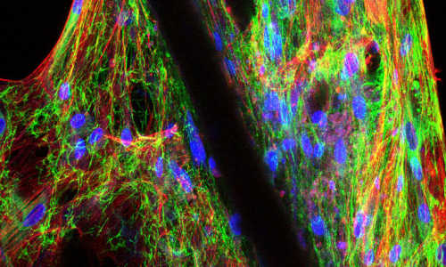
“The morphology and the structural organisation of the artificial organ are extremely similar to if not indistinguishable from a natural skeletal muscle,” says Cesare Gargioli of the University of Rome, one of the lead authors of the study.

In future, irreversibly damaged muscles could be restored by implanting the patient’s own cells within the hydrogel matrix on top of a residual muscle, adjacent to the damaged area. “While we are encouraged by the success of our work in growing a complete intact and functional mouse leg muscle we emphasize that a mouse muscle is very small and scaling up the process for patients may require significant additional work,” comments EMBO Member Giulio Cossu, one of the authors of the study. The next step in the work will be to use larger animal models to test the efficacy of this approach before starting clinical studies.

Story Source:

The above story is based on materials provided by EMBO – excellence in life sciences.

Share14Tweet9Share2ShareShareShare2

Related Posts

Mini breasts grown in petri dishes for cancer research

June 12, 2015

Soft-tissue engineering for hard-working cartilage

May 16, 2015

Breakthrough in 3-D printing of replacement body parts

April 29, 2015

New material for creating artificial blood vessels

April 28, 2015
Please login to join discussion

POPULAR NEWS

  • Enhancing Spiritual Care Education in Nursing Programs

    157 shares
    Share 63 Tweet 39
  • Robotic Ureteral Reconstruction: A Novel Approach

    81 shares
    Share 32 Tweet 20
  • Digital Privacy: Health Data Control in Incarceration

    63 shares
    Share 25 Tweet 16
  • PTSD, Depression, Anxiety in Childhood Cancer Survivors, Parents

    149 shares
    Share 60 Tweet 37

About

We bring you the latest biotechnology news from best research centers and universities around the world. Check our website.

Follow us

Recent News

Linking Critical Thinking to EBP Adoption in Nursing

Hemodynamic States of PDA Affect Preterm Organ Perfusion

Massachusetts Prescription Overdose Deaths: A 2000-2023 Study

Subscribe to Blog via Email

Success! An email was just sent to confirm your subscription. Please find the email now and click 'Confirm' to start subscribing.

Join 72 other subscribers
  • Contact Us

Bioengineer.org © Copyright 2023 All Rights Reserved.

Welcome Back!

Login to your account below

Forgotten Password?

Retrieve your password

Please enter your username or email address to reset your password.

Log In
No Result
View All Result
  • Homepages
    • Home Page 1
    • Home Page 2
  • News
  • National
  • Business
  • Health
  • Lifestyle
  • Science

Bioengineer.org © Copyright 2023 All Rights Reserved.