• HOME
  • NEWS
  • EXPLORE
    • CAREER
      • Companies
      • Jobs
    • EVENTS
    • iGEM
      • News
      • Team
    • PHOTOS
    • VIDEO
    • WIKI
  • BLOG
  • COMMUNITY
    • FACEBOOK
    • INSTAGRAM
    • TWITTER
Monday, November 10, 2025
BIOENGINEER.ORG
No Result
View All Result
  • Login
  • HOME
  • NEWS
  • EXPLORE
    • CAREER
      • Companies
      • Jobs
        • Lecturer
        • PhD Studentship
        • Postdoc
        • Research Assistant
    • EVENTS
    • iGEM
      • News
      • Team
    • PHOTOS
    • VIDEO
    • WIKI
  • BLOG
  • COMMUNITY
    • FACEBOOK
    • INSTAGRAM
    • TWITTER
No Result
View All Result
Bioengineer.org
No Result
View All Result
Home NEWS Science News Biology

Scientists find mechanism behind precise spinal cord development

Bioengineer by Bioengineer
June 29, 2017
in Biology
Reading Time: 2 mins read
0
Share on FacebookShare on TwitterShare on LinkedinShare on RedditShare on Telegram
IMAGE

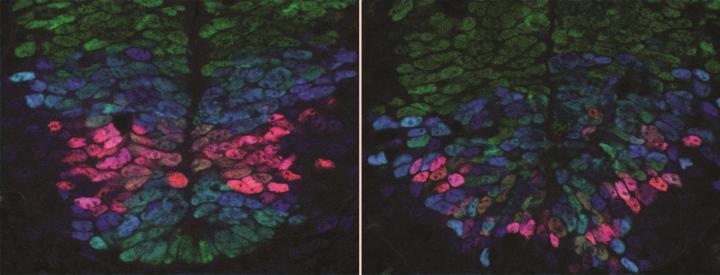
Credit: Anna Kicheva

Scientists have uncovered how nerve cells in the spinal cord are organised in precise patterns during embryo development – a finding that could give insight into regenerative medicine.

As embryos grow and develop they need the right cell types to end up in the right places inside forming organs. This is particularly important in the spinal cord where different nerve cell types must be accurately positioned so that circuits can assemble properly to control muscle movement. But until now the mechanism underlying nerve cell organisation in the spinal cord has remained poorly understood.

In a study published in Science, researchers at the Francis Crick Institute, the Institute of Science and Technology (Austria) and Ecole Polytechnique Fédérale de Lausanne (Switzerland) report that cells destined to become nerve cells in developing mouse embryos use two different signals spreading from opposite sides of the spinal cord – the back and belly side – to measure their position accurately. Based on this map, they turn into the appropriate nerve cell type. The research was funded by the European Research Council and Wellcome.

The team of biologists, physicists and engineers found that the amounts of the two signals originating from the back and belly sides of the body affect gene activity in developing nerve cells. Based on this gene activity in early development, the cells turn into the appropriate nerve cell type for that position in the spinal cord.

"We've made an important step in understanding how the diverse cell types in the spinal cord of a developing embryo are organised in a precise spatial pattern. The quantitative measurements and new experimental techniques we used, as well as the combined effort of biologists, physicists and engineers were key. This allowed us to gain new insight into the exquisite accuracy of embryonic development and revealed that cells have remarkable ability of to orchestrate precise tissue development," says Anna Kicheva, Group Leader at IST Austria.

"We have shed light on the long-standing question of how developing tissues produce the right cells in the right place in the right numbers," says James Briscoe, Group Leader at the Francis Crick Institute. "It's likely that similar strategies are used in other developing tissues and our findings might be relevant to these cases. In the long run this will help inform the use of stem cells in approaches such as tissue engineering and regenerative medicine. However, there is still much more to learn and we need to continue developing these interdisciplinary collaborations to further our biological understanding."

The paper 'Decoding of position in the developing neural tube from antiparallel morphogen gradients' is published in Science.

###

Media Contact

Greta Keenan
[email protected]
020-379-65252
@thecrick

http://www.crick.ac.uk

############

Story Source: Materials provided by Scienmag

Share12Tweet7Share2ShareShareShare1

Related Posts

TFAP2C Boosts CST1, Promoting Breast Cancer Growth

TFAP2C Boosts CST1, Promoting Breast Cancer Growth

November 10, 2025
blank

Decoding Cell Type and State Through Feature Selection

November 10, 2025

Embryonic Heat Manipulation: Metabolic Programming Insights

November 9, 2025

ProteinFormer: Transforming Protein Localization with Bioimages

November 9, 2025
Please login to join discussion

POPULAR NEWS

  • blank

    Stinkbug Leg Organ Hosts Symbiotic Fungi That Protect Eggs from Parasitic Wasps

    315 shares
    Share 126 Tweet 79
  • ESMO 2025: mRNA COVID Vaccines Enhance Efficacy of Cancer Immunotherapy

    207 shares
    Share 83 Tweet 52
  • New Study Suggests ALS and MS May Stem from Common Environmental Factor

    139 shares
    Share 56 Tweet 35
  • Sperm MicroRNAs: Crucial Mediators of Paternal Exercise Capacity Transmission

    1303 shares
    Share 520 Tweet 325

About

We bring you the latest biotechnology news from best research centers and universities around the world. Check our website.

Follow us

Recent News

TFAP2C Boosts CST1, Promoting Breast Cancer Growth

Impact of Music vs. Storytelling on Preterm Infants

New Study Empowers Eczema Patients to Decide Their Own Bathing Frequency

Subscribe to Blog via Email

Enter your email address to subscribe to this blog and receive notifications of new posts by email.

Join 70 other subscribers
  • Contact Us

Bioengineer.org © Copyright 2023 All Rights Reserved.

Welcome Back!

Login to your account below

Forgotten Password?

Retrieve your password

Please enter your username or email address to reset your password.

Log In
No Result
View All Result
  • Homepages
    • Home Page 1
    • Home Page 2
  • News
  • National
  • Business
  • Health
  • Lifestyle
  • Science

Bioengineer.org © Copyright 2023 All Rights Reserved.