
In a groundbreaking study published in the esteemed journal Radiology, researchers have unveiled six distinct breast parenchymal texture patterns that may signal an increased risk of developing breast cancer. This large-scale investigation leverages advanced radiomic techniques applied to mammographic images, marking a significant leap forward in breast cancer risk stratification beyond traditional breast density measures.
Breast density has long been recognized as a critical factor in assessing cancer risk. Dense breast tissue, characterized by a predominance of glandular and fibrous tissue rather than fat, complicates cancer detection because both dense tissue and tumors appear white on conventional mammograms. This chromatic similarity makes early malignancies more challenging to identify, potentially delaying diagnosis and treatment. However, not all dense breasts are alike; the microstructural patterns within the tissue vary considerably, which may harbor clues about an individual’s susceptibility to cancer.
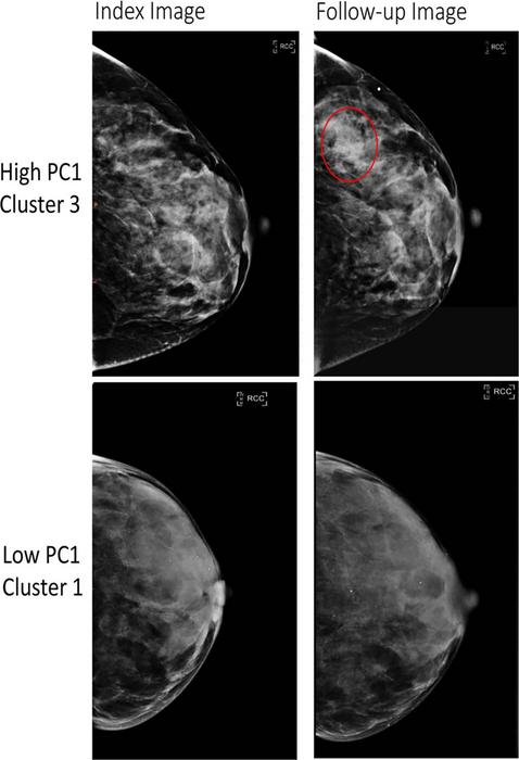
The study, spearheaded by a multidisciplinary team including epidemiologists and radiologists, analyzed over 30,000 mammograms from women with no prior history of breast cancer, drawn from three diverse screening cohorts. Utilizing radiomics—a cutting-edge computational approach that extracts and quantifies intricate patterns from medical images invisible to the naked eye—the researchers identified 390 quantitative imaging features. These features were then distilled into six prominent phenotypes or texture patterns that epitomize variations in breast parenchymal architecture.
To validate their findings, the team examined these phenotypes in an independent cohort exceeding 3,500 women, including those who later developed invasive breast cancer and those who remained cancer-free. Strikingly, the existence of specific radiomic phenotypes correlated strongly with an elevated risk of invasive disease. These associations persisted across racial lines, providing a robust framework for predicting cancer risk with potential implications for personalized screening programs.
One of the most compelling revelations was the apparent differential impact of these radiomic phenotypes among Black compared to white women. The phenotypes demonstrated a starker association with breast cancer risk in Black women—a population historically burdened with more aggressive cancer subtypes and poorer outcomes. This disparity underscores the urgency of integrating novel imaging biomarkers into risk models tailored for diverse populations to mitigate existing health inequities.
Beyond risk prediction, the phenotypes also showed promise in forecasting diagnostic challenges such as false-negative mammograms—where cancer lesions are missed during routine screening—and interval cancers, which are diagnosed between scheduled mammograms and often have worse prognoses. Being able to anticipate these diagnostic blind spots could revolutionize follow-up protocols and preventive interventions.
Dr. Celine M. Vachon, a senior author and professor of epidemiology at the Mayo Clinic, emphasized the transformative potential of this research. She noted that discerning subtle tissue textural differences offers a more nuanced understanding of breast biology and individual risk, transcending the binary dense vs. non-dense paradigm. This nuanced stratification may allow clinicians to tailor screening intervals and supplemental imaging strategies, optimizing early detection while minimizing unnecessary procedures.
Co-senior author Despina Kontos from Columbia University highlighted the imperative to focus on populations disproportionately affected by aggressive breast cancers. The discovery that radiomic phenotypes may capture risk disparities reinforces the role of advanced imaging analytics in driving equitable healthcare solutions. Incorporating such phenotypes alongside genetic and lifestyle factors could refine risk prediction algorithms and empower precision medicine.
The study also opens avenues for exploring these texture phenotypes in three-dimensional mammography (tomosynthesis) or other imaging modalities, potentially enhancing detection accuracy. By integrating radiomic data with genomic and clinical variables, future research could foster comprehensive risk profiles that fundamentally alter breast cancer prevention and early detection.
Karla M. Kerlikowske, co-senior author and professor at the University of California San Francisco, remarked on the clinical significance of identifying women at greatest risk of invasive and aggressive cancers. Early identification facilitates timely interventions which may reduce morbidity and mortality, and potentially diminish treatment intensity, benefiting both patients and healthcare systems.
This research exemplifies how artificial intelligence and data-driven imaging analysis are transforming radiology. By quantifying features invisible to human assessment, radiomics demands a reevaluation of conventional screening metrics and represents an extraordinary leap toward personalized, predictive oncology.
The integration of radiomic phenotypes into existing clinical workflows promises to enhance breast cancer risk models and screening efficiency. These findings underscore a future where breast cancer screening is tailored not only by age and density, but also by subtle architectural tissue signatures predictive of cancer risk.
Looking ahead, the authors intend to expand their studies to larger, more diverse populations within the United States, investigate the potential added value of three-dimensional mammographic imaging, and assess the synergistic impact of combining imaging phenotypes with genetic profiling and lifestyle data. This comprehensive approach aspires to delineate more accurately who is truly at increased risk for invasive breast cancer and who may safely benefit from less frequent surveillance.
In summary, this landmark study harnesses the power of radiomics to decode breast tissue texture, revealing phenotypic signatures associated with differential cancer risk. By illuminating these subtle imaging biomarkers, researchers have paved the way for refined, equitable, and personalized breast cancer screening and prevention strategies that could save countless lives.
Subject of Research: People
Article Title: Radiomic Parenchymal Phenotypes of Breast Texture from Mammography and Association with Risk of Breast Cancer
News Publication Date: 13-May-2025
Web References:
Radiology Journal
Radiological Society of North America (RSNA)
RadiologyInfo.org
Image Credits: Radiological Society of North America (RSNA)
Keywords: Breast carcinoma, Mammography, Cancer
Tags: advanced radiology researchbreast cancer risk factorsbreast density and cancer diagnosisbreast parenchymal texture patternscomputational imaging in medicineepidemiology of breast cancermammographic imaging techniquesmicrostructural patterns in breast tissuemultidisciplinary approaches to cancer researchradiomics in cancer detectionrisk stratification in oncologyscreening methods for breast cancer
