• HOME
  • NEWS
  • EXPLORE
    • CAREER
      • Companies
      • Jobs
    • EVENTS
    • iGEM
      • News
      • Team
    • PHOTOS
    • VIDEO
    • WIKI
  • BLOG
  • COMMUNITY
    • FACEBOOK
    • INSTAGRAM
    • TWITTER
Tuesday, July 7, 2026
BIOENGINEER.ORG
No Result
View All Result
  • Login
  • HOME
  • NEWS
  • EXPLORE
    • CAREER
      • Companies
      • Jobs
        • Lecturer
        • PhD Studentship
        • Postdoc
        • Research Assistant
    • EVENTS
    • iGEM
      • News
      • Team
    • PHOTOS
    • VIDEO
    • WIKI
  • BLOG
  • COMMUNITY
    • FACEBOOK
    • INSTAGRAM
    • TWITTER
  • HOME
  • NEWS
  • EXPLORE
    • CAREER
      • Companies
      • Jobs
        • Lecturer
        • PhD Studentship
        • Postdoc
        • Research Assistant
    • EVENTS
    • iGEM
      • News
      • Team
    • PHOTOS
    • VIDEO
    • WIKI
  • BLOG
  • COMMUNITY
    • FACEBOOK
    • INSTAGRAM
    • TWITTER
No Result
View All Result
Bioengineer.org
No Result
View All Result
Home NEWS Science News Biology

Scientists discover how proteins form crystals that tile a microbe’s shell

Bioengineer by Bioengineer
December 17, 2019
in Biology
Reading Time: 3 mins read
0
IMAGE
Share on FacebookShare on TwitterShare on LinkedinShare on RedditShare on Telegram

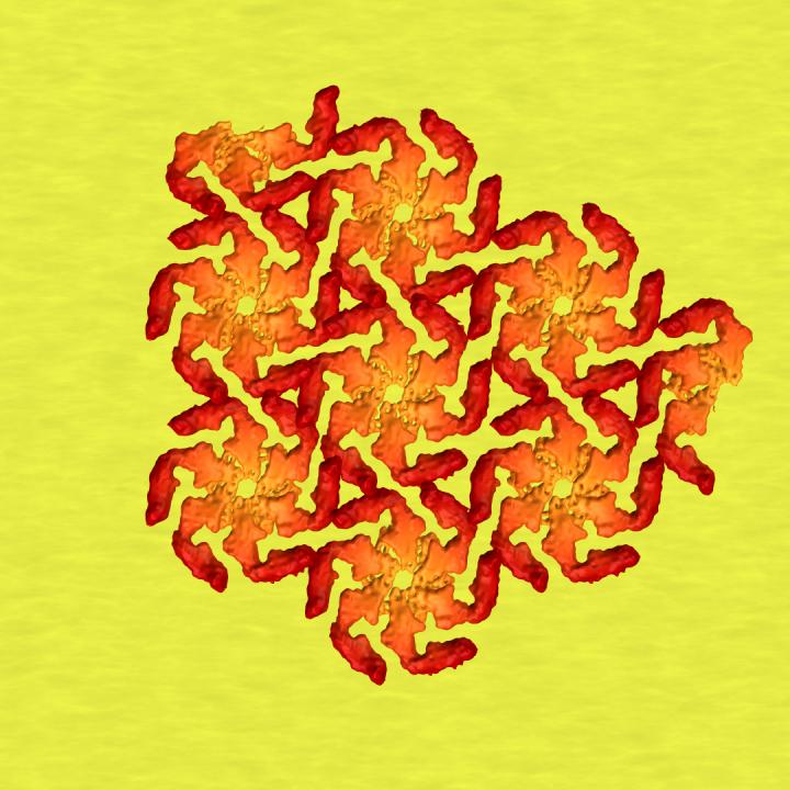
A new understanding of the nucleation process could shed light on how the shells help microbes interact with their environments, and help people design self-assembling nanostructures for various tasks.
IMAGE

Credit: Greg Stewart/SLAC National Accelerator Laboratory


Many microbes wear beautifully patterned crystalline shells, which protect them from a harsh world and can even help them reel in food. Studies at the Department of Energy’s SLAC National Accelerator Laboratory and Stanford University have revealed this food-reeling process and shown how shells assemble themselves from protein building blocks.

Now the same team has zoomed in on the very first step in microbial shell-building: nucleation, where squiggly proteins crystallize into sturdy building blocks, much like rock candy crystallizes around a string dipped into sugar syrup.

The results, published today in the Proceedings of the National Academy of Sciences, could shed light on how the shells help microbes interact with other organisms and with their environments, and also help scientists design self-assembling nanostructures for various tasks.

Jonathan Herrmann, a graduate student in Professor Soichi Wakatsuki’s group at SLAC and Stanford, collaborated with the structural molecular biology team at SLAC’s Stanford Synchrotron Radiation Lightsource (SSRL) on the study. They scattered a powerful beam of X-rays off protein molecules that were floating in a solution to see how the atomic structures of the molecules changed as they nucleated into crystals. Meanwhile, other researchers made a series of cryogenic electron microscope (cryo-EM) images at various points in the nucleation process to show what happened over time.

They found out that crystal formation takes place in two steps: One end of the protein molecule nucleates into crystal while the other end, called the N-terminus, continues to wiggle around. Then the N-terminus joins in, and the crystallization is complete. Far from being a laggard, the N-terminus actually speeds up the initial nucleation step ­- although exactly how it does this is still unknown, the researchers said – and this helps explain why microbial shells can form so quickly and efficiently.

###

Some of the X-ray data was collected at Lawrence Berkeley National Laboratory’s Advanced Light Source, which like SSRL is a DOE Office of Science user facility. Researchers from the University of British Columbia and from Professor Lucy Shapiro’s laboratory at Stanford also contributed to this work, funded in part by the National Institute of General Medical Sciences and the Chan Zuckerberg Biohub. Work in Wakatsuki’s labs at SLAC and Stanford was funded by a Laboratory Directed Research and Development grant from SLAC, the DOE Office of Science’s Office of Biological and Environmental Research, and Stanford’s Precourt Institute for Energy.

Citation: Jonathan Herrmann et al., PNAS, 17 December 2019 (10.1073/pnas.1909798116)

SLAC is a vibrant multiprogram laboratory that explores how the universe works at the biggest, smallest and fastest scales and invents powerful tools used by scientists around the globe. With research spanning particle physics, astrophysics and cosmology, materials, chemistry, bio- and energy sciences and scientific computing, we help solve real-world problems and advance the interests of the nation.

SLAC is operated by Stanford University for the U.S. Department of Energy’s Office of Science. The Office of Science is the single largest supporter of basic research in the physical sciences in the United States and is working to address some of the most pressing challenges of our time.

Media Contact
Glennda Chui
[email protected]
510-507-2766

Original Source

https://www6.slac.stanford.edu/news/2019-12-17-scientists-discover-how-proteins-form-crystals-tile-microbes-shell.aspx

Related Journal Article

http://dx.doi.org/10.1073/pnas.1909798116

Tags: BiologyMicrobiology
Share12Tweet8Share2ShareShareShare2

Related Posts

Here are a few rewritten headlines for a science magazine post, each with a slightly different tone: Intriguing & poetic: How do organs sculpt themselves? Sea stars hold the secret Direct & research-focused: Sea stars reveal the hidden rules of organ formation Metaphorical & inviting: Tiny architects beneath the waves: What sea stars teach us about building organs Short & punchy: Star-shaped clues to how our organs take shape Question-led: Could a sea star show us how organs form? Elegant & feature-style: The body’s blueprint, glimpsed in a sea star’s arm

July 6, 2026
Bacteria evolve faster with unconventional gene copies — Biology

Bacteria evolve faster with unconventional gene copies

July 6, 2026

Neighbours rewire soil feedback via root microbiome shifts

July 6, 2026

Evolution-Inspired Biosensors Revolutionize Lipid Tracking in Real Time

July 2, 2026
Please login to join discussion

POPULAR NEWS

  • Detection of EDCs in Breast Milk and Infant Urine Up to Six Months Highlights Early Exposure Risks

    77 shares
    Share 31 Tweet 19
  • New Drug Candidate Developed at McMaster Shows Potential for Treating Brain Cancer

    58 shares
    Share 23 Tweet 15
  • Saying Goodbye to PGY-6: Pediatric Fellowship Realities

    103 shares
    Share 41 Tweet 26
  • KTU Researchers Explore Ultrasound’s Role in Enhancing Blood Flow Beyond Diagnostics

    53 shares
    Share 21 Tweet 13

About

We bring you the latest biotechnology news from best research centers and universities around the world. Check our website.

Follow us

Recent News

Flame retardant BDE-209 targets molecularly linked to ulcerative colitis

Ultra-high frequency particle impacts mimic rockbursts to shatter hard rock

Kidney transplant outcomes in older adults studied by German researchers

Subscribe to Blog via Email

Enter your email address to subscribe to this blog and receive notifications of new posts by email.

Join 83 other subscribers
  • Contact Us

Bioengineer.org © Copyright 2023 All Rights Reserved.

Welcome Back!

Login to your account below

Forgotten Password?

Retrieve your password

Please enter your username or email address to reset your password.

Log In
No Result
View All Result
  • Homepages
    • Home Page 1
    • Home Page 2
  • News
  • National
  • Business
  • Health
  • Lifestyle
  • Science

Bioengineer.org © Copyright 2023 All Rights Reserved.