
Credit: Aleksandr Mishin
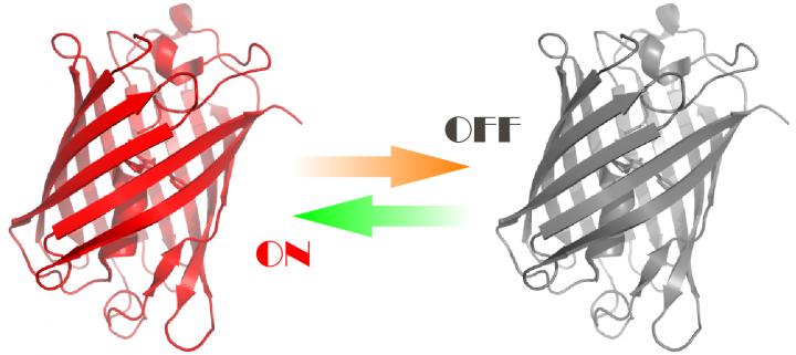
Researchers developed fluorescent proteins that can be controlled by orange and green light. These proteins will help to study processes in living cells. The work was supported by Russian Science Foundation (RSF) grant, and the results were published in Nature Methods.
Fluorescent proteins emit intense visible light with wavelengths ranging from 390 to 700 nm. Natural functions of such proteins are rather diverse: for instance, some species of jellyfish use green fluorescent spots to attract various small organisms that serve as food. Optical properties of certain fluorescent proteins can be controlled with light. For instance, such proteins can be "turned on" or "turned off" therefore they are called switchable. Switchable fluorescent proteins are widely used in a new group of methods called super-resolution fluorescence microscopy (nanoscopy), which allow of extremely detailed intracellular structures imaging. Scientists usually use blue or violet irradiation for such microscopy, which is very toxic for cells as it disrupts their normal physiology and can even cause death.
"We were the first to create photoswitchable fluorescent proteins with optical properties that can be controlled using green and orange light, rather than blue-violet radiation. The advantage of this is minimal harm to cells. We used new proteins to observe cytoskeleton changes in living cells over time," explained Aleksandr Mishin, Ph.D, Senior Researcher of the Shemyakin-Ovchinnikov Institute of Bioorganic Chemistry of the Russian Academy of Sciences, who headed up the RSF project.
In order to create such fluorescent proteins, the scientists altered them by directed and random mutagenesis using a method called polymerase chain reaction. After that, the scientists cloned proteins and selected the best-performing ones using a microscope. The authors analyzed the results of experiments carried out by other biologists and found out how the microenvironment of the chromophore (the aromatic amino acid residue responsible for light absorption in the protein) must be altered to make it capable of photoswitching.
However, the expected outcome does have its side effects: for instance, the brightness of the protein is reduced. Now the researchers use random mutagenesis to find additional mutations, which compensate for side effects while preserving the target mutation.
The proteins developed are called reporter proteins, as they act as "spies" within cells. They are attached to other proteins, which can then be traced in a living cell. The detailed information obtained thereby can be used in basic science or biomedical research. For instance, tumor cells in cancer patients exhibit dramatic disturbances of cell mobility and dynamic structural changes in the cytoskeleton, a carcass in the cytoplasm of a living cell. Meanwhile, the investigation of these processes in living cells by nanoscopy is difficult due to the overly intense irradiation of samples, making it necessary to use methods that are less toxic for the organism.
The authors used their development to carry out RESOLFT super-resolution microscopy. The proteins have an important feature: their photoswitching is very efficient, meaning that fluorescence can be switched on and off in milliseconds. This does not suit all microscopy methods. In some cases, the high speed will only be a nuisance. In RESOLFT, the on-off cycle is repeated many times for adjacent points that are scanned with laser beams. The better the switching time of a fluorescent tag, the faster the complete image can be obtained, as photoswitching at each point requires less time.
"The fluorescent proteins we created will enable super-resolution microscopy without harming the living cell, which opens up opportunities for studying dynamic processes within the cell," Aleksandr Mishin concluded.
###
The work was carried out in collaboration with scientists from the Royal Institute of Technology in Sweden and Albert Einstein College of Medicine in the United States.
Media Contact
Alexander Mishin
[email protected]
http://www.akson.science
Related Journal Article
http://dx.doi.org/10.1038/s41592-018-0052-9
