In the evolving landscape of pediatric radiology, advancements in imaging techniques are paramount for enhancing diagnostic accuracy and safety. One of the latest innovations making waves in the field is the integration of dual-energy computed tomography (DECT) virtual monoenergetic images (VMIs) for evaluating pediatric hand angiography. This technique presents a significant leap in imaging technology, merging high-resolution imaging with the subtle nuances necessary for pediatric anatomy.
The research led by Xu, Liu, and Xu explores the application of DECT VMIs specifically tailored for use in pediatric cases involving hand angiography. The study meticulously examines how dual-energy techniques can improve the clarity and detail of angiographic images, especially in younger patients whose anatomy differs considerably from adults. By harnessing the unique properties of DECT, this research aims to refine diagnostic processes while minimizing the exposure to ionizing radiation, a critical consideration in pediatric medicine.
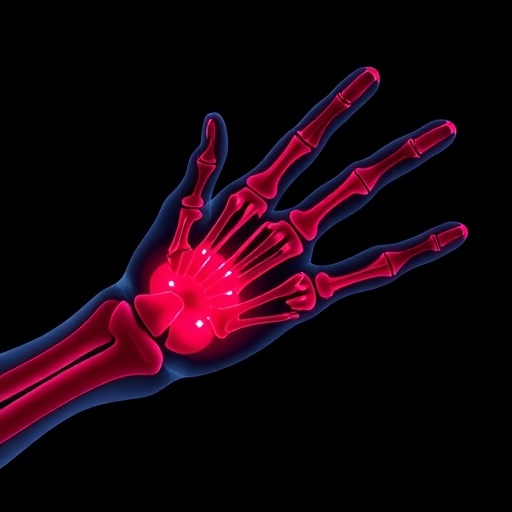
Traditional angiography techniques, although effective, often require higher doses of radiation, posing risks to a child’s developing tissues and organs. In contrast, DECT employs two different energy levels to acquire images, allowing for the differentiation of materials based on their attenuation characteristics. This capability is particularly beneficial in identifying vascular structures and potential abnormalities within the intricate network of blood vessels in a child’s hand, where subtle variations can significantly influence treatment decisions.
The implications of this research extend beyond mere imaging. The ability to produce virtual monoenergetic images enhances contrast resolution without increasing radiation dose, which is a crucial factor in pediatric radiology. VMIs can effectively reduce motion artifacts commonly seen in younger patients who may find it challenging to remain still during imaging procedures, thus yielding higher quality images with potentially lower repeat rates.
In addition to providing more detailed anatomical visualization, the application of DECT VMIs also facilitates improved differentiation between vascular phases. This ensures that radiologists and clinicians can observe blood flow dynamics in real-time, potentially identifying vascular malformations such as arteriovenous malformations or vascular tumors with unprecedented accuracy. The study’s findings highlight how this technology enhances the overall diagnostic confidence among pediatric radiologists, which is vital for formulating effective treatment plans.
As the field progresses toward personalized medicine, the significance of advanced imaging techniques like DECT VMIs becomes increasingly apparent. The research underscores the importance of employing imaging modalities that not only enhance diagnostic potential but also prioritize patient safety and comfort. Such advancements are essential in a pediatric setting, where the stakes are particularly high due to the vulnerability of young patients.
Through rigorous clinical trials and examinations, the researchers have provided substantial evidence supporting the adoption of DECT VMIs in routine practice. Their findings are expected to prompt shifts in imaging protocols across pediatric hospitals, advocating for the integration of this technology as a standard practice rather than an adjunct. This could lead to widespread improvements in patient health outcomes, empowering clinicians with more reliable imaging options.
Moreover, as medical imaging technology continues to evolve, the emphasis on training radiologists to proficiently interpret DECT VMI results is crucial. The knowledge of how to utilize and interpret these images will define the next generation of pediatric radiologists. This study serves not only as a groundbreaking contribution to current medical literature but also as a guide for educational institutions in refining their training programs around advanced imaging techniques.
For medical imaging enthusiasts and professionals alike, this research opens the door to a more nuanced understanding of how dual-energy computed tomography can transform pediatric angiography. It stands as a testament to the collaborative efforts of researchers aiming to bridge gaps in pediatric care through technological advancements. The excitement surrounding this study reflects a broader trend in medicine, where the focus is increasingly shifting toward harnessing technology for concrete improvements in patient care.
As the medical community actively seeks ways to reduce radiation exposure while maximizing diagnostic precision, the insights provided by this research could herald a new standard in how pediatric hand angiography is performed. The anticipation surrounding widespread implementation hints at a future where children receive safer and more accurate imaging, ultimately leading to better clinical outcomes. The researchers hope that their findings will inspire further studies that continue to explore the full potential of dual-energy CT imaging across various anatomical regions and clinical scenarios.
The quest for perfection in imaging remains unending, but the strides made in this study serve as a beacon of progress. As clinicians and radiologists embrace this innovative approach, the potential to revolutionize pediatric care comes into sharper focus. Ultimately, as we move toward a future defined by precision and safety in medicine, the value of studies like this cannot be overstated, ensuring that technological advancements translate directly into improved care for the youngest and most vulnerable patients in our healthcare systems.
Subject of Research: Pediatric hand angiography using dual-energy computed tomography.
Article Title: Application value of dual-energy computed tomography virtual monoenergetic images for pediatric hand angiography.
Article References:
Xu, H., Liu, B., Xu, Z. et al. Application value of dual-energy computed tomography virtual monoenergetic images for pediatric hand angiography.
Pediatr Radiol (2026). https://doi.org/10.1007/s00247-026-06524-2
Image Credits: AI Generated
DOI: https://doi.org/10.1007/s00247-026-06524-2
Keywords: Pediatric Radiology, Dual-Energy Computed Tomography, Virtual Monoenergetic Images, Angiography, Imaging Technology, Radiation Safety, Diagnostic Imaging.
Tags: DECT virtual monoenergetic imagesdiagnostic accuracy in childrendual-energy computed tomographyevaluating pediatric vascular structureshigh-resolution imaging techniquesinnovative imaging technologiesionizing radiation safetyminimizing radiation exposurepediatric anatomy imaging challengespediatric hand angiographypediatric radiology advancementsvascular imaging in pediatrics


