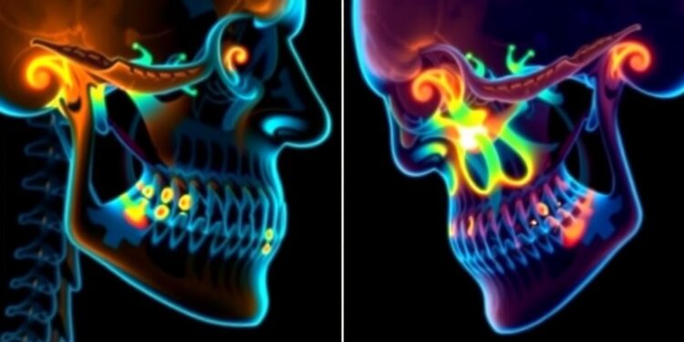

A groundbreaking study has opened new avenues in understanding the complex mechanisms of temporomandibular joint (TMJ) disorders, providing insights that could significantly enhance the treatment landscape for millions affected by this debilitating condition. Utilizing a state-of-the-art in vivo imaging technology, researchers captured real-time functional activity within mouse models afflicted by TMJ injury and inflammation. This innovative approach not only broadens the current understanding of TMJ pain pathways but also holds promise for the development of more effective therapeutic interventions.
The research, recently published in the esteemed journal Pain, was spearheaded by Dr. Yu Shin Kim, an associate professor in the Department of Oral and Maxillofacial Surgery at The University of Texas Health Science Center at San Antonio (UT Health San Antonio). The study marked a significant milestone by revealing the simultaneous activity of over 3,000 trigeminal ganglion neurons, which play a crucial role in transmitting sensory information from the face, mouth, and head to the brain. This detailed neuronal activity was meticulously observed, allowing researchers to explore how these nerves respond to various stimuli, particularly in the context of injury or inflammation associated with TMJ disorders.
Dr. Kim emphasized the revolutionary nature of their imaging technique, stating, “With our novel imaging technique and tools, we can see each individual neuron’s activity, pattern, and dynamics while also visualizing the ensemble network of over 3,000 neurons in real-time.” This groundbreaking capability allows for an unprecedented examination of the neuronal networks that underpin TMJ pain, paving the way for insights that may lead to more sophisticated pain management strategies.
Temporomandibular joint disorders rank as the second most prevalent musculoskeletal disorders in the United States, affecting up to 12% of the population. The condition often manifests as chronic facial pain, with many patients experiencing inadequate relief from existing treatment modalities. This underscores the urgency for in-depth research into the underlying pathology of TMJ disorders, which could explain why current therapies frequently yield unsatisfactory results for patients.
The study revealed a startling connection between TMJ injuries and the subsequent inflammatory processes that ensue within the joint. Researchers demonstrated that injury or misalignment of the TMJ instigates inflammation that can propagate to associated nerve networks, culminating in heightened and chronic pain. Notably, findings indicated that over 100 neurons could fire simultaneously in response to the joint’s nociceptive signals, elucidating specific neural pathways that are integral to the experience of TMJ-related pain.
Furthermore, the research explored the interconnection between TMJ disorders and other syndromes, such as migraines and chronic headaches. This correlation aligns with previous literature suggesting that TG neuron inflammation can lead to pain referral to proximate regions, including those that are classically associated with migraine disorders. Understanding these overlapping pain mechanisms further emphasizes the need for comprehensive studies of TMJ disorders as multifaceted conditions that may share neurophysiological origins with other chronic pain syndromes.
A particularly notable focus of the study was the investigation into Calcitonin Gene-Related Peptide (CGRP) antagonists as a potential treatment modality for TMJ pain. CGRP is a neuropeptide known to be involved in inflammatory processes and pain transmission, and its elevated levels have been documented in patients suffering from TMJ disorders. Remarkably, Dr. Kim’s research team uncovered that reducing CGRP levels in the synovial fluid of affected joints led to significant pain alleviation, alongside a decrease in hypersensitivities observed in trigeminal ganglion neurons.
Despite the lack of FDA-approved treatments that specifically target TMJ disorders, the promising results of this research indicate that CGRP antagonists—previously sanctioned for migraine therapy—may serve as viable candidates for addressing TMJ pain. This finding not only sheds light on an innovative therapeutic pathway but also highlights the broader implications such advancements may have on the treatment of various chronic pain conditions.
The study stands as a pivotal juncture in the quest to unravel the complexities of TMJ disorders, providing deeper insights into the neuronal mechanisms that govern pain perception and chronicity. The implications of this research extend beyond TMJ disorders alone, potentially guiding future strategies for pain management in a variety of other chronic conditions. Dr. Kim articulated the team’s overarching vision: “Our hope is that this approach will not only advance treatments for TMJ disorders but also pave the way for understanding and managing various chronic pain conditions more effectively.”
The potential impact of these discoveries cannot be overstated. Advancements in in vivo imaging techniques empower researchers to visualize and interrogate pain at its source—meaning clinicians can ultimately transition toward more targeted and personalized approaches for pain relief. As the medical community seeks to comprehend and rectify the complexities of chronic pain syndromes, studies such as this drive a much-needed discourse about the future of pain management.
In concluding remarks, the considerable advances presented in this research beckon a reevaluation of current treatment paradigms for TMJ disorders. With an increased understanding of neural activity and inflammation, medical professionals will be better equipped to address the underlying factors contributing to TMJ pain and its associated comorbidities. This research not only delivers hope to patients but also lays the groundwork for future exploration into the uncharted territories of pain etiology and management.
Subject of Research: Temporomandibular Joint Disorders and Pain Mechanisms
Article Title: Elucidation of neuronal activity in mouse models of temporomandibular joint injury and inflammation by in vivo GCaMP Ca2+ imaging of intact trigeminal ganglion neurons
News Publication Date: December 2024
Web References: Study, UT Health San Antonio News
References: DOI 10.1097/j.pain.0000000000003421
Image Credits: UT Health San Antonio
Keywords: In vivo imaging, Chronic inflammation, Neuroscience, TMJ disorders, Pain management, CGRP antagonists, Trigeminal ganglion neurons, Musculoskeletal disorders, Pain transmission, Neuronal pathways.
Tags: chronic pain management strategieschronic TMJ pain treatmentscutting-edge imaging techniques in medicineDr. Yu Shin Kim research findingsin vivo imaging technologymechanisms of TMJ inflammationoral and maxillofacial surgery advancementsreal-time imaging in neurosciencetherapeutic interventions for TMJTMJ disorders researchtrigeminal ganglion neurons activityunderstanding TMJ pain pathways

