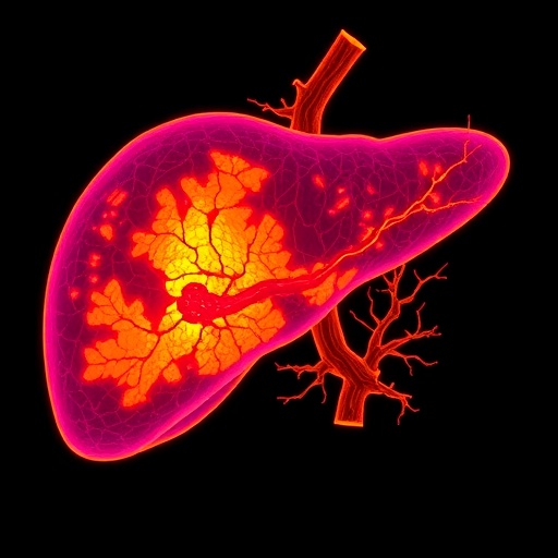
In an era where artificial intelligence and medical imaging are increasingly interwoven, a groundbreaking study has emerged that promises to redefine approaches to liver segmentation in diagnostic imaging. The researchers, led by Debnath, Rahman, and Azam, have developed a pioneering framework known as FSS-ULivR (Few-Shot Segmentation for Unifying Liver Representation), which significantly enhances clinical outcomes in liver imaging. Their work, as published in the esteemed Journal of Cancer Research and Clinical Oncology, addresses a pressing need for improved image segmentation techniques in a field where precision and efficiency are paramount.
The core of the FSS-ULivR framework revolves around the concept of few-shot learning—this paradigm allows the model to learn from a remarkably small number of annotated imaging examples. Traditional machine learning techniques typically require extensive datasets to achieve reliable performance, often posing hurdles in the medical imaging field where labeled data can be scarce. FSS-ULivR not only surmounts this challenge but elevates the process of liver imaging to unprecedented levels, facilitating better diagnostic accuracy while simultaneously conserving time and resources.
At the heart of this innovative framework lies a dual approach combining unified representations and sophisticated attention mechanisms. Unified representations enable the model to create a comprehensive understanding of the liver’s anatomical structures, which is crucial for effective segmentation. By leveraging representations that encompass various imaging modalities—such as CT scans and MRI—the researchers ensure that their framework is robust across different technologies, making it adaptable and versatile in diverse clinical settings.
Attention mechanisms play a pivotal role in honing the performance of FSS-ULivR. These mechanisms allow the model to prioritize critical features within an image, effectively simulating a human-like focus that enhances the segmentation process. Through this advanced technique, the model can discern between the liver and surrounding tissues and pathologies with remarkable precision. The application of attention mechanisms in medical imaging is a salient leap towards bridging the gap between artificial intelligence capabilities and clinical expertise.
A significant benefit of adopting FSS-ULivR is its ability to operate effectively in environments where traditional models might fail. Many existing segmentation methods falter when presented with atypical or varied datasets; however, the few-shot learning approach allows FSS-ULivR to generalize better, even with limited training data. The implications of this are vast, particularly in cases where patients may present with unique anatomical features or pathologies that deviate from the norm. The potential for widespread application in diverse patient populations could lead to a significant advancement in personalized medicine.
Further enhancing the framework’s utility is its adaptability to ongoing advancements in image acquisition technologies. As medical imaging continues to evolve, with new methodologies and modalities being introduced, FSS-ULivR’s unified representation approach means that it can adapt to these changes without necessitating extensive retraining. This characteristic ensures that the framework remains relevant and continues to provide value in a fast-paced technological landscape.
Moreover, the researchers embarked on rigorous evaluations of FSS-ULivR’s performance against standard benchmarks in liver segmentation. The results were not only statistically significant but also showcased improvements in segmentation accuracy that could translate into tangible clinical benefits. Increased accuracy can lead to better treatment planning, reduced surgical risks, and improved patient outcomes—all critical factors in the realm of oncology.
As the healthcare industry moves towards integrated care solutions, the implementation of advanced segmentation frameworks such as FSS-ULivR becomes crucial. This is particularly true in multidisciplinary settings where radiologists, oncologists, and surgeons must collaborate closely to ensure comprehensive patient care. Enhanced liver imaging through improved segmentation enhances communication among these teams, facilitating a more streamlined decision-making process.
Healthcare institutions considering the adoption of FSS-ULivR are likely to benefit from not only enhanced image analysis but also improved workflow efficiency. With quicker and more accurate segmentation, healthcare professionals can devote more time to interpreting results and devising patient-centric treatment plans rather than spending excessive time on image processing. This efficiency gain could have notable implications for reducing overall healthcare costs while enhancing the quality of care delivered to patients.
Moreover, the implications of FSS-ULivR extend beyond immediate clinical applications. Its development exemplifies the potential for innovative algorithms to drive advancements in the broader field of medical imaging. The fusion of few-shot learning with advanced attention mechanisms sets the stage for future research endeavors aimed at tackling various challenges within medical imaging domains. By inspiring subsequent studies, FSS-ULivR contributes to the continuous advancement of knowledge, promoting an era of ongoing innovation.
In the realm of education, the framework exemplifies a paradigm shift that can influence training methodologies for upcoming medical professionals. As medical imaging techniques evolve, the need for modern educational curricula that incorporate such advanced frameworks becomes paramount. The integration of FSS-ULivR into training programs could equip future radiologists and oncologists with the skills necessary to leverage state-of-the-art technology effectively, culminating in better-prepared healthcare practitioners.
In conclusion, the FSS-ULivR framework emerges as a transformative force in liver imaging, heralding a new era for precision healthcare. It encapsulates the synthesis of few-shot learning principles and advanced attention mechanisms, paving the way for better segmentation outcomes in a clinical setting. As the medical community continues to explore innovative technologies and methodologies, it is evident that FSS-ULivR represents a crucial step toward advancing liver imaging and, by extension, improving patient care across the globe.
The research conducted by Debnath, Rahman, and Azam underscores the importance of continuous innovation in medical technology. With a future that holds the promise of even more groundbreaking advancements, FSS-ULivR stands as an emblem of how artificial intelligence can be harnessed to revolutionize medical practices and enhance patient outcomes, thus fulfilling the long-standing quest for precision in medicine.
Subject of Research: Liver segmentation in medical imaging
Article Title: FSS-ULivR: a clinically-inspired few-shot segmentation framework for liver imaging using unified representations and attention mechanisms
Article References:
Debnath, R.K., Rahman, M.A., Azam, S. et al. FSS-ULivR: a clinically-inspired few-shot segmentation framework for liver imaging using unified representations and attention mechanisms. J Cancer Res Clin Oncol 151, 215 (2025). https://doi.org/10.1007/s00432-025-06256-0
Image Credits: AI Generated
DOI:
Keywords: Few-shot learning, liver segmentation, medical imaging, attention mechanisms, artificial intelligence, cancer diagnosis, unified representations, clinical outcomes
Tags: advanced liver representation frameworkartificial intelligence in diagnosticsattention mechanisms in segmentationclinical outcomes in liver diagnosticsfew-shot learning in medical imagingFSS-ULivR frameworkinnovative approaches to diagnostic imagingJournal of Cancer Research and Clinical Oncologyliver imaging segmentationmachine learning for medical imagingprecision in liver imagingresource-efficient medical imaging techniques
