
Credit: Lei Xi, University of Electronic Science and Technology of China
WASHINGTON — For the first time, researchers have followed the development of blood vessels in zebrafish embryos without using any labels or contrast agents, which may disturb the biological processes under study.
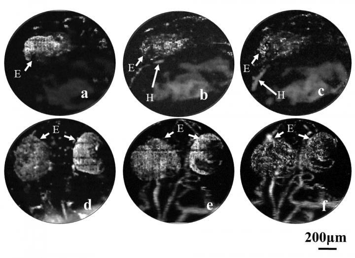
"We successfully visualized the development of the cardio-cerebrovascular system in the early embryonic stage of zebrafish," said Lei Xi of the University of Electronic Science and Technology of China. "Since the zebrafish remains one of the most important models to study human diseases, we believe that our work is important to understanding both brain disorders and cardiovascular diseases in humans."
Zebrafish embryos are transparent, making them an excellent model for researchers who want to study how vertebrate organisms develop and grow. But until now, visualizing biological processes with high-resolution optical imaging techniques such as confocal fluorescence microscopy and two-photon microscopy has typically required a fluorescence label or contrast agent. Getting these agents into a zebrafish is not only complex and time consuming, but can sometimes change the natural physiological processes taking place in the zebrafish.
In The Optical Society (OSA) journal Biomedical Optics Express, Xi and colleagues report using optical resolution photoacoustic microscopy (ORPAM) for label-free imaging of vessel development over the whole body of a zebrafish embryo with the technique's low-resolution mode. They also zoomed in to follow the formation of the vessels associated with the heart and brain by using the high-resolution mode.
Using sound to make images
ORPAM doesn't require any contrast agents because it forms images by detecting ultrasonic waves created when molecules absorb light. This relatively new microscopy technique can provide three-dimensional images with a penetration depth and spatial resolution comparable to two-photon microscopy.
In the new study, the researchers used a transmission-mode ORPAM system with a low-resolution objective to image the vessel development at various time points for the whole zebrafish with 3.5-micron resolution. They also conducted ORPAM with a high-resolution objective to follow the development of the brain and heart vasculature with a resolution of 1.5 microns. The images revealed the exact timing of heart vessel development as well as details of how whole-body vasculature develops in the zebrafish.
"The ability to visualize dynamic development during the embryonic stage, when the blood vessels initially form and develop, brings the potential to use zebrafish to investigate various human cardiovascular and cerebrovascular diseases in the future," said Xi. The approach could be useful for studying epilepsy, heart disease, hardening and narrowing of the arteries, high blood pressure, heart attacks, strokes and blood clots in the brain.
The researchers are working to improve the imaging speed of the method, which currently requires 25 minutes for a whole-body zebrafish embryo scan. They would also like to combine ORPAM with new gene expression techniques to study the genetic causes of vascular diseases.
###
Paper: Q. Chen, T. Jin, W. Qi, X. Mo, L. Xi, "Label-free photoacoustic imaging of the cardio-cerebrovascular development in the embryonic zebrafish," Biomed. Opt. Express, Volume 8, Issue 4, 2359-2367 (2017). DOI: 10.1364/BOE.8.002359.
About Biomedical Optics Express
Biomedical Optics Express is OSA's principal outlet for serving the biomedical optics community with rapid, open-access, peer-reviewed papers related to optics, photonics and imaging in the life sciences. The journal scope encompasses theoretical modeling and simulations, technology development, and biomedical studies and clinical applications. It is published by The Optical Society and edited by Christoph Hitzenberger, Medical University of Vienna. Biomedical Optics Express is an open-access journal and is available at no cost to readers online at OSA Publishing.
About The Optical Society
Founded in 1916, The Optical Society (OSA) is the leading professional organization for scientists, engineers, students and business leaders who fuel discoveries, shape real-life applications and accelerate achievements in the science of light. Through world-renowned publications, meetings and membership initiatives, OSA provides quality research, inspired interactions and dedicated resources for its extensive global network of optics and photonics experts. For more information, visit osa.org/100.
Media Contacts:
Rebecca B. Andersen
The Optical Society
[email protected]
+1 202.416.1443
Joshua Miller
The Optical Society
[email protected]
+1 202.416.1435
Media Contact
Joshua Miller
[email protected]
202-416-1435
@opticalsociety
http://www.osa.org
############
Story Source: Materials provided by Scienmag


