Researchers at Rutgers Health have unveiled crucial insights into the aggressive lung disease known as idiopathic pulmonary fibrosis (IPF). This innovative study sheds light on the role of specialized immune cells, particularly misplaced plasma cells, that not only proliferate but may actively drive the progression of this devastating illness. The findings offer hope for novel treatment paths for a disease that is notoriously lethal, with a staggering 80% of patients succumbing within a decade of diagnosis.
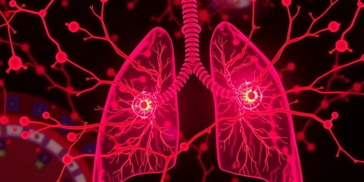
Idiopathic pulmonary fibrosis is characterized by the accumulation of scar tissue within the lungs, resulting in progressively compromised respiratory function. The relentless scarring impedes the transfer of oxygen, leaving patients struggling for breath and facing perilous limitations in their daily lives. While a few drugs have been approved to address IPF, they yield only marginal benefits, leaving many patients with limited therapeutic options. Furthermore, lung transplantation, while potentially life-saving for some, comes with its own set of risks, including a grim 50% mortality rate within five years post-transplant.
The research team employed advanced spatial mapping techniques to critically analyze lung tissues harvested from both healthy individuals and those suffering from fatal IPF. In a startling revelation, they uncovered a significant proliferation of plasma cells in the fibrotic lung regions of IPF patients. Plasma cells, which primarily reside in bone marrow, are critical in producing antibodies. Traditionally, their presence in lung tissues is minimal, yet in the case of IPF, these cells appear to be over-represented.
Professor Qi Yang of Rutgers University, a co-author of the study, emphasized the startling contrast between healthy lungs and those afflicted with IPF. He noted that in normal lung tissues, plasma cells are nearly absent, while in the tissue afflicted with IPF, these antibody-producing cells dominate the scarring regions. This aberration raises significant questions about the immune response mechanisms contributing to the pathology of IPF.
In pursuit of understanding the underlying cellular networks governing this abnormal immune reaction, the researchers made groundbreaking observations. They identified unique mural cells encircling blood vessels, which actively produce signaling proteins critical for orchestrating immune responses. Additionally, they documented previously unidentified fibroblasts, a type of cell known for its role in tissue scarring, that secrete compounds attracting plasma cells to the sites of lung damage.
Dr. Reynold Panettieri, another senior author of the research, highlighted the uniqueness of the fibroblast population identified. While fibroblasts have been well documented as key players in fibrosis across various organs—including skin and brain—the distinctive population of fibroblasts found in the lungs appears previously uncharacterized. This finding not only underscores the complexity of lung pathology in IPF but also offers potential for targeted therapies.
Encouraged by their findings concerning plasma cell accumulation, the research team proceeded to test therapeutic strategies using live mice. Through experiments designed to inhibit the signaling pathways integral to plasma cell localization in the lungs, they successfully demonstrated a reduction in plasma cell numbers and corresponding mitigation of lung scarring. Such targeted treatments could prove revolutionary in slowing the progression of IPF among human patients.
Interestingly, existing medications designed to combat plasma cell diseases, such as multiple myeloma, may hold promise for repurposing in the treatment of IPF. Dr. Yang speculated that if plasma cells are indeed responsible for producing detrimental antibodies, it may be imperative to eliminate them from the lungs, thereby preventing the chronic immune responses that exacerbate the disease.
Prior studies have highlighted a correlation between elevated antibody levels in the lungs of IPF patients and their autoimmune responses. This new research elucidates the origins of these antibodies and illustrates how plasma cells, under abnormal conditions, accumulate within the lung tissue. As such, the study lays the groundwork for exploring potential autoimmune links to IPF.
The mechanisms by which these antibodies inflict tissue damage are multifaceted. The Rutger’s researchers suggest that antibody-antigen complexes may mediate the production of transforming growth factor-beta from pulmonary macrophages, a cytokine known to propagate fibrotic processes. By establishing a clearer connection between immune dysregulation and lung tissue pathology, this research may guide the development of more effective interventions.
In light of these findings, the research team has positioned themselves to further investigate the intriguing possibility of autoantibodies generated by plasma cells targeting healthy lung tissue. They plan to delve deeper into understanding how mural cells and fibroblasts redefine their characteristics and functions in the context of IPF. This comprehensive approach could be critical as they continue to unravel the underlying pathology of this life-threatening disease.
The collaboration behind this research draws expertise from both the Child Health Institute of New Jersey and the Rutgers Institute for Translational Medicine and Science, indicating a concerted effort that bridges animal model research with careful analysis of human end-stage lung tissues. Such interdisciplinary approaches are vital for the advancement of our understanding and treatment of complex diseases like IPF.
Given the alarmingly high mortality rate associated with IPF, the implications of this research are profound. It offers a newly illuminated target for therapy, potentially allowing for more focused interventions that could significantly improve outcomes in affected patients. As this research progresses, it may pave the way for transformative medical strategies aimed at countering the relentless advance of idiopathic pulmonary fibrosis.
In conclusion, the revelations regarding misplaced immune cell networks in IPF not only enhance our understanding of the disease’s pathology but also inspire hope for new, effective therapeutic strategies. With a strong potential for drug repurposing and innovative treatments stemming from this study, researchers remain optimistic about impacting the lives of patients suffering from this debilitating condition. As research continues, the exploration of the autoimmune connections in IPF may unlock further breakthroughs in treatment modalities, heralding a new dawn for those affected by this difficult ailment.
Subject of Research: Animals
Article Title: Distinct mural cells and fibroblasts promote pathogenic plasma cell accumulation in idiopathic pulmonary fibrosis
News Publication Date: 20-Feb-2025
Web References: European Respiratory Journal
References: DOI: 10.1183/13993003.01114-2024
Image Credits: N/A
Keywords: Idiopathic pulmonary fibrosis, plasma cells, immune response, fibroblasts, autoimmunity, lung disease, treatment options.
Tags: advanced spatial mapping in medical researchchallenges in treating pulmonary fibrosisidiopathic pulmonary fibrosis researchimmune cell networks in lung diseaseinnovative treatments for lung fibrosisinsights into fatal lung diseaseslung scarring and respiratory functionlung transplantation risks and outcomesnovel therapeutic pathways for IPFprognosis for IPF patientsrole of plasma cells in IPFRutgers Health lung disease study


