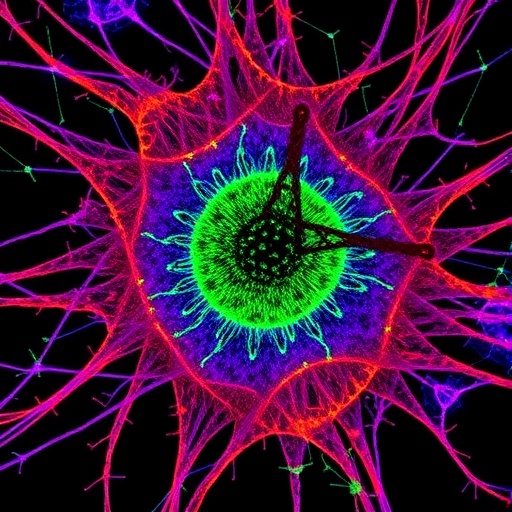
In a groundbreaking advancement for cancer diagnostics and therapeutic strategies, researchers from the Institute of Hygiene at the University of Münster have unveiled a novel analytical method that merges fluorescence microscopy with MALDI-2 mass spectrometry imaging. This innovative approach unlocks unprecedented insight into the minute chemical landscapes of tumor tissues at a single-cell level, promising a paradigm shift in how oncologists understand tumor microenvironments and cellular interactions. Published in the esteemed journal Nature Communications, this research paves the way for more rapid, precise diagnoses and personalized treatments, fundamentally enhancing the prospects for patient outcomes.
Understanding the microscopic interplay among cells within tumors is crucial for effective cancer treatment. Tumors comprise a complex ecosystem where cancer cells interact dynamically with surrounding stromal cells and infiltrating immune cells. Such interactions often dictate tumor growth, metastasis, and response to therapy. While fluorescence microscopy has long been able to characterize cell types through specific protein biomarkers, it has lacked the capacity to map intricate chemical profiles within the same spatial context. The newly developed technique overcomes this limitation by integrating fluorescence imaging directly with matrix-assisted laser desorption/ionisation (MALDI) mass spectrometry, enabling the correlation of cellular identity with their unique metabolic signatures.
Matrix-assisted laser desorption/ionisation, or MALDI, operates by using a laser to ionize molecules from tissue samples, which are then identified and quantified based on their mass-to-charge ratios within a mass spectrometer. The primary challenge of traditional MALDI has been its sensitivity and spatial resolution limits, both critical for single-cell analysis. The Münster team’s approach incorporates the advanced MALDI-2 technique, employing a secondary laser for post-ionisation that significantly amplifies the detection sensitivity for various small molecules, lipids, and metabolites critical to tumor biology. This dual-laser setup is combined with transmission mode geometry, whereby the laser irradiates the tissue from the opposite side, substantially enhancing spatial resolution down to about one micrometer.
What truly sets this methodology apart is the direct integration of a fluorescence microscope within the same mass spectrometry instrument. This configuration allows for simultaneous fluorescence-based cell identification and mass spectrometric chemical profiling on the exact same tissue sections, with no need for tissue relocation or re-preparation. By optimizing the sample preparation protocols to be compatible with both fluorescence markers and mass spectrometry requirements, the team has established a seamless workflow that preserves both molecular and cellular integrity.
The ability to precisely identify cell types through fluorescence signals corresponding to specific proteins and subsequently map their complex metabolomic and lipidomic profiles within the spatial context of tissue opens new investigative avenues. For example, researchers can now observe subtle metabolic differences not only between cancerous and non-cancerous cells but also among neighboring tumor cells with distinct phenotypes. This fine-grained chemical imaging lays the foundation for deciphering the biochemical dialogues within tumor microenvironments—information that has been largely inaccessible until now.
Moreover, by visualizing previously hidden metabolic heterogeneity, the technique illuminates mechanisms of tumor progression and immune evasion. The interplay between malignant cells and immune infiltrates is a key determinant of whether cancer remains localized or spreads. Understanding these chemical interactions can reveal novel biomarkers indicative of aggressive tumor behavior or susceptibility to immunotherapies. As Dr. Alexander Potthoff, the study’s first author, emphasizes, this capability marks the first occasion where cell types can be directly matched with their chemical signatures in situ, offering unprecedented insights into cellular communication.
The technical innovation relies heavily on the use of an inverse irradiation geometry — transmission mode — which was previously described but not yet combined with MALDI-2 and integrated fluorescence microscopy in this manner. The transmission mode facilitates laser focus through the sample itself rather than from above, refining the laser spot size and thereby enhancing spatial resolution critical for single-cell analysis. The MALDI-2 secondary laser then further ionizes desorbed molecules, bolstering sensitivity across a broad range of chemical classes including lipids and metabolites that are otherwise challenging to detect.
This multiplexed analytical platform is poised to benefit diverse fields beyond oncology, including cell biology, immunology, and tumor biology research. Established fluorescence microscopy techniques can be complemented and augmented by adding chemical context, enabling deeper functional studies into cellular metabolism, signaling pathways, and microenvironmental influences. Furthermore, the clinical potential is immense. The method could be adapted for rapid biopsy assessment in clinical workflows, providing clinicians with more comprehensive information to guide treatment choices with higher precision.
The researchers foresee further technical refinements enhancing spatial resolution into the sub-micron scale — approaching a few hundred nanometers. Such advancements would unlock the capacity to chemically analyze intracellular organelles such as lipid droplets, vesicles, or synaptic structures within cells, vastly expanding the granularity of spatial biology. This could accelerate novel drug discovery, revealing targets previously hidden within the complex chemical architecture of cells and tissues, ultimately driving more effective therapies.
This pioneering work also highlights close collaboration between academia and industry, involving the University of Münster and Bruker Daltonics in Bremen. The synergy between fundamental research expertise and industrial instrumentation innovation underscores how cross-sector partnerships can stimulate technical breakthroughs with translational potential. Financial backing by the German Research Foundation (DFG) was instrumental in bringing this vision to fruition.
Overall, this integrated fluorescence microscopy–t-MALDI-2 mass spectrometry imaging platform represents a transformative leap forward by bridging molecular imaging and spatial biology at single-cell resolution. Such capabilities not only deepen fundamental understanding of cancer biology but also herald future clinical tools that could revolutionize diagnostic and therapeutic pathways. As researchers continue to refine and apply this technology, the outlook for personalized medicine and targeted cancer therapies grows ever brighter.
Subject of Research:
Integration of fluorescence microscopy with MALDI-2 mass spectrometry imaging for single-cell metabolic profiling in tumor tissues.
Article Title:
Spatial biology using single-cell mass spectrometry imaging and integrated microscopy
News Publication Date:
15-Oct-2025
Web References:
http://dx.doi.org/10.1038/s41467-025-64603-8
Image Credits:
Peter Leßmann
Keywords:
Cancer diagnostics, single-cell imaging, MALDI mass spectrometry, MALDI-2, fluorescence microscopy, tumor microenvironment, metabolomics, lipidomics, spatial biology, transmission mode, mass spectrometry imaging, integrated microscopy
Tags: cancer cell dynamicscancer treatment personalizationcellular metabolic signatureschemical signals in tumorsfluorescence microscopy and MALDI imaginginnovative cancer research techniquesmass spectrometry in oncologysingle-cell cancer diagnosticstherapeutic strategies for cancertumor microenvironment analysistumor-stromal cell interactions


