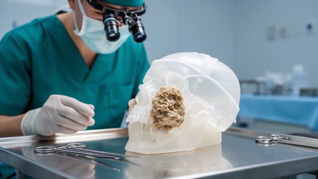
A 25-year-old woman walked into an oral and maxillofacial surgery clinic with a painless swelling on the left side of her face that had been slowly growing for about five years. Imaging revealed a mass measuring roughly 7 by 6 by 2 centimeters, centered on her left anterior maxillary alveolus and extending upward into the maxillary sinus until it reached the level of the orbital floor. Despite its enormous size, the tumor had displaced rather than invaded the structures around it, lifting the orbital floor and shifting the medial maxillary wall while leaving vision, eye movement, and facial sensation completely intact. The case, reported in Clinical Case Reports, describes how surgeons removed the entire lesion in one piece through a conservative approach that spared the patient a partial maxillectomy and preserved one of her front teeth.
The mass was a central ossifying fibroma, or COF, a benign but potentially expansile tumor belonging to the fibro-osseous family of craniofacial lesions. These conditions are characterized by the replacement of normal bone with cellular fibrous tissue containing variable amounts of mineralized material, including woven and lamellar bone and cementum-like deposits. The three main entities seen in clinical practice are fibrous dysplasia, cemento-osseous dysplasia, and ossifying fibroma, and they share striking histological similarities while differing fundamentally in pathophysiology, biological behavior, and treatment. Because of this overlap, a reliable diagnosis requires the combination of clinical, radiographic, and microscopic observations rather than histology alone, since a true neoplasm may demand complete excision while a dysplastic lesion may only warrant observation or contouring.
COF itself is considered a benign tumor with autonomous neoplastic progression, which distinguishes it from fibrous dysplasia, a lesion that typically stabilizes once skeletal maturation is complete. The World Health Organization reinstated the term cemento-ossifying fibroma in the fourth edition of its classification for lesions arising in the tooth-bearing regions of the jaws, reflecting a suggested connection to the periodontal ligament and its multipotent mesenchymal cells, which can produce bone, cementum, and fibrous tissue. The tumor shows a clear female predilection, with reported female-to-male ratios of roughly 2:1 to 5:1, and is most commonly diagnosed in the second to fourth decades of life. The mandible is by far the most frequently affected site: one reported series documented mandibular involvement in 93 percent of 28 cases, while another recorded posterior mandibular involvement in 55.5 percent of 61 patients. Lesions arising in the anterior maxilla are especially uncommon, and extension into the sinonasal tract and maxillary sinus is rarer still, with only a handful of cases described in multicenter and specialized rhinological studies.
The anatomy of the maxilla helps explain how such a tumor can reach enormous dimensions before anyone notices. The pneumatized maxillary sinus provides a relatively low-resistance space into which an expanding lesion can grow, potentially delaying presentation until facial asymmetry, nasal obstruction, dental displacement, or orbital involvement appears. In this patient, computed tomography showed a distinct, expansile lesion with heterogeneous internal density, a mixture of hypodense and hyperdense components compatible with progressive mineralization. The anterior, lateral, and posterior walls of the maxillary antrum were noticeably thinned and expanded, but there was no obvious cortical destruction and no extraosseous soft-tissue mass. The lesion deviated the nasal septum to the right, restricted the left nasal airway, and made direct contact with the left orbital floor, elevating it despite significant thinning and displacement. Crucially, there was no periosteal erosion, orbital fat infiltration, or extraocular muscle involvement.
The clinical presentation was equally striking for what it lacked. The patient reported no pain, paresthesia, discharge, epistaxis, epiphora, diplopia, or visual impairment, and she had no history of radiation therapy, prior surgery, or facial trauma. Blood tests ruled out an underlying metabolic disorder, with serum calcium, phosphate, alkaline phosphatase, and parathyroid hormone all within reference ranges, excluding hyperparathyroidism-jaw tumor syndrome. Extraoral examination showed significant left facial asymmetry with elevation of the upper lip and effacement of the nasolabial fold, while intraoral findings included a bony-hard, non-tender swelling of the left anterior maxilla with labial and palatal expansion, obliteration of the vestibule, and displacement of both central and lateral incisors. The differential diagnosis ranged from benign fibro-osseous lesions and odontogenic tumors to low-grade osteosarcoma and other sinonasal malignancies, but the prolonged indolent course, preserved cortical boundaries, and absence of soft-tissue infiltration strongly favored a benign process.
Three surgical options were weighed: an endoscopic transnasal technique, conservative total enucleation, and a broad partial maxillectomy with reconstruction. Partial maxillectomy would have sacrificed the anterior maxillary alveolus and surrounding teeth, creating substantial reconstructive morbidity, while an endoscopic approach was deemed inappropriate because the lesion originated in the anterior maxillary alveolus rather than the sinonasal cavity. After thorough discussion, the team chose conservative enucleation of the entire lesion, relying on the clearly defined radiographic borders and an apparent surgical plane around the tumor. Under general anesthesia, they elevated a full-thickness mucoperiosteal flap, removed a thin eggshell-like cortical layer covering the tumor, and developed a distinct cleavage plane with blunt periosteal dissection. The tumor was delivered as a single solid mass measuring 7 by 6 by 2 centimeters, avoiding piecemeal excision. When the orbital floor was examined afterward, it was intact but noticeably thinned and displaced; the periorbita remained intact and no orbital reconstruction was necessary.
The decision about the adjacent teeth was made according to remaining osseous support. The left central incisor had lost its supporting bony housing entirely and was extracted, but sufficient labial and palatal bone remained around the lateral incisor to permit its preservation. Histopathological examination of the specimen showed irregular trabeculae of woven and lamellar bone prominently rimmed by plump osteoblasts within a moderately to highly cellular fibrous stroma of bland spindle-shaped fibroblasts, along with focal cementum-like spherules. No nuclear pleomorphism, atypical mitotic activity, permeative bone infiltration, or malignant features were observed. The team paid particular attention to excluding the juvenile ossifying fibroma variants, which can behave more aggressively and recur more often after conservative surgery. Juvenile trabecular ossifying fibroma is characterized by thin, anastomosing strands of immature osteoid, which were absent here, and juvenile psammomatoid ossifying fibroma features dense, widespread psammomatoid ossicles and typically involves sinonasal and orbital bones, neither of which matched this lesion. Taken together with the patient’s age and slow five-year course, the findings supported a conventional central ossifying fibroma.
The postoperative course was uneventful, with no significant hemorrhage, wound dehiscence, infection, oro-antral communication, orbital complication, or infraorbital paraesthesia. Facial symmetry improved substantially, the upper lip returned to a normal resting position, and nasal airflow improved, while ocular position, extraocular movements, and visual acuity remained normal. The patient was enrolled in a structured clinical and radiological monitoring program with evaluations at 1, 3, 6, and 12 months. At the one-year mark she remained asymptomatic, the preserved lateral incisor was still vital, firm, and functional, and prosthetic replacement of the extracted central incisor had been completed. A 12-month radiographic evaluation showed increasing osseous regeneration of the surgical cavity, a preserved orbital floor, restored maxillary sinus aeration, and no evidence of recurrent or residual disease.
The authors emphasize that this favorable outcome should be regarded as a case-specific observation rather than a general treatment guideline. Conservative enucleation was achievable only because of a well-defined, intact capsule and a distinct dissection plane; in poorly marginated lesions with aggressive or infiltrative growth, radical resection is generally indicated. Published recurrence rates for COF vary substantially, from 6.7 percent in a large series managed predominantly by curettage to 12 percent in a systematic review, and up to 23 percent in maxillary and sinonasal lesions and 28.6 percent in pediatric jaw lesions, reflecting differences in subtype, location, surgical technique, completeness of excision, and follow-up duration. The main limitation of the report is the 12-month follow-up, which cannot establish long-term tumor control because recurrence may occur later, and a single case cannot compare conservative enucleation with radical resection. Nevertheless, the case suggests that tumor size and sinonasal-orbital extension alone may not require radical surgery in conventional COF when a clear cleavage plane exists, and that surgical planning should focus on the lesion’s relationship to vital structures and the feasibility of complete extracapsular excision, with longer follow-up and additional cases needed to confirm the durability of this approach.
Subject of Research: Conservative surgical management of a giant central ossifying fibroma of the anterior maxilla with maxillary sinus and orbital floor extension
Article Title: Giant Central Ossifying Fibroma of the Anterior Maxilla and Maxillary Sinus With Orbital Floor Extension: A Case Report
Article References: Alashwal, A., Rajeh, N., Alkherbash, A. M., & Madfa, A. (2026). Giant Central Ossifying Fibroma of the Anterior Maxilla and Maxillary Sinus With Orbital Floor Extension: A Case Report. Clinical Case Reports, 14(11), Article e73803. https://doi.org/10.1002/ccr3.73803
Image Credits: AI Generated
DOI: 10.1002/ccr3.73803
Keywords: central ossifying fibroma, maxilla, maxillary sinus, orbital floor, fibro-osseous lesion, enucleation, maxillectomy, oral and maxillofacial surgery, case report, juvenile ossifying fibroma, recurrence rate, CT imaging
News Source: Ophelia Keating. (October 28, 2026). Research report: Giant Central Ossifying Fibroma of the Anterior Maxilla and Maxillary Sinus With Orbital Floor Extension: A Case Report. Scienmag.