Credit: Toubibe/Pixabay
Universidad Carlos III de Madrid (UC3M) is participating in a research project together with the Hospital General Universitario Gregorio Marañón (HGUGM), the Instituto de Investigación Sanitaria San Carlos and the company Sedecal Molecular Imaging (SMI), project coordinator, to develop a new high-precision radiology system for coronavirus pulmonary involvement.
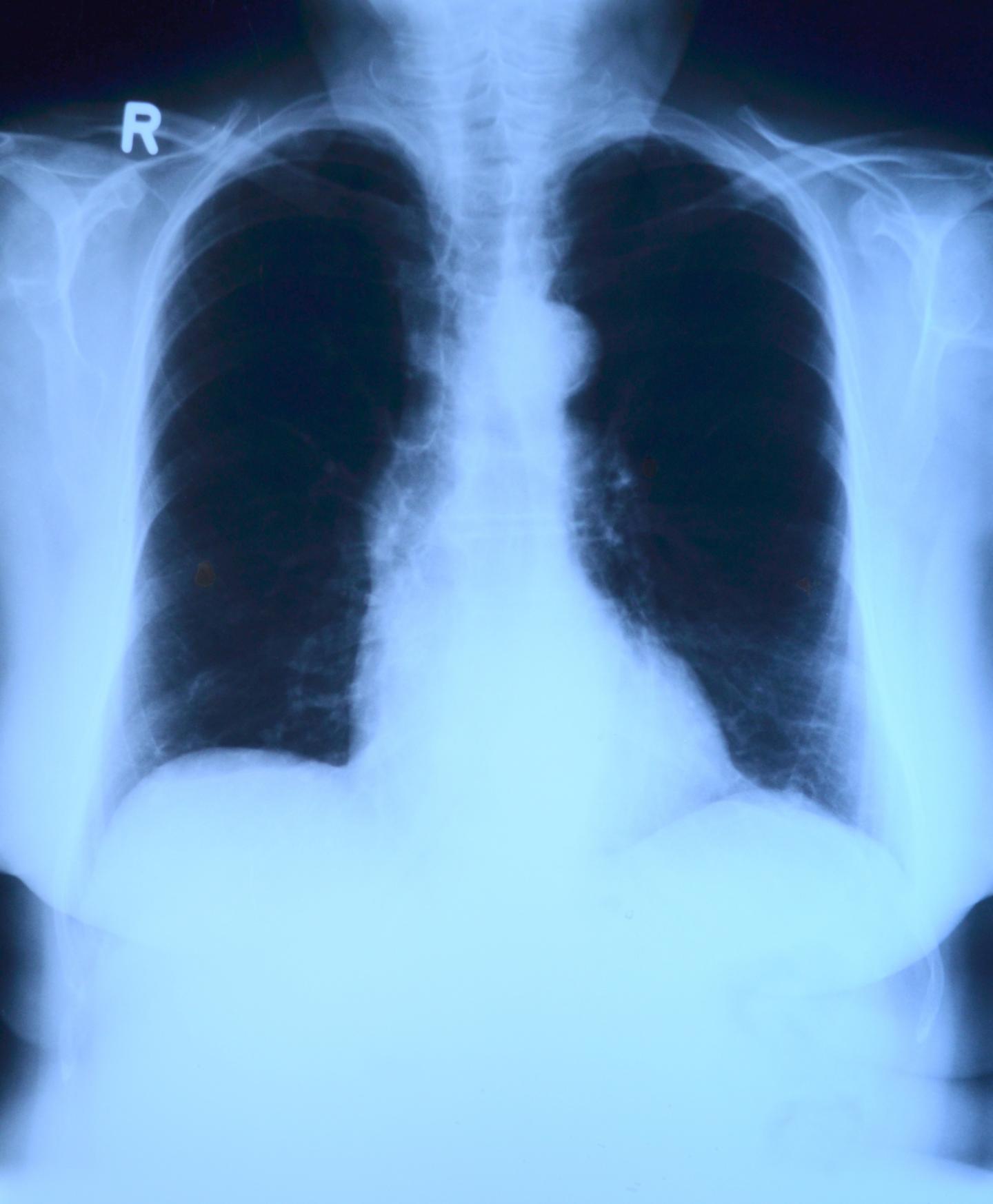
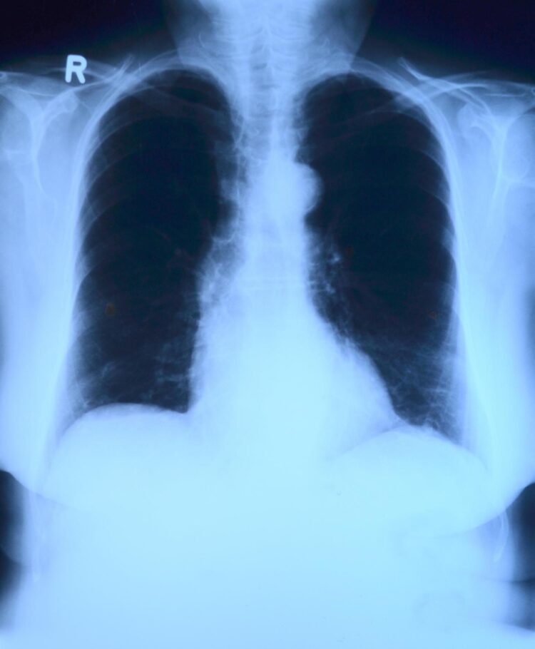
Radiology constitutes a key element in the management of patients suffering from COVID-19 because it enables decision making in processes such as hospital admission, type of treatment and ICU transfer. The x-ray films currently used are not very sensitive and underestimate pulmonary damage if compared with results from a CAT (computerized axial topography) scan. However, it is not viable to use the latter test on all patients with suspected COVID-19 because of issues regarding equipment availability and logistics.
Within the framework of this project, the researchers seek to combine Artificial Intelligence (AI) and Tomosynthesis (with low dose x-ray imaging)to develop a low cost device able to substantially increase the diagnostic accuracy of the radiology, up to levels comparable to that of a CAT scan. Furthermore, this system would offer much greater availability and would be more versatile, and could even be installed in vehicles or in portable triage tents.
“The incorporation of IA algorithms can contribute to facilitating diagnosis, accelerating image analysis and reducing the dose of radiation the patient receives”, explained the project’s principal investigator, Manuel Desco, the researcher from the Bioengineering and Aerospace Engineering Department at UC3M and the Instituto de Investigación Sanitaria Gregorio Marañón at HGUGM.
The general objective of the initiative is to adapt technological developments already on course to meet the specific needs of radiology imaging required by the COVID-19 epidemic. The project is estimated to last six months, and when finished, its aim is to have a new completely functional radiology system, capable of making “quasi-tomographic” X-ray studies.
This system has numerous advantages over current approaches. First, it allows greater diagnostic accuracy in radiology, which is critical for the clinical management of the patient. Secondly, it increases availability of diagnostic tools at an affordable cost. Third, it facilitates the epidemiological characterization of the outbreak, improving detection of pulmonary involvement. Fourth, it simplifies the work of the radiologists. Finally, it guarantees optimization of the dose administered to the patient.
###
Media Contact
Javier Alonso
[email protected]
Original Source
https:/


