• HOME
  • NEWS
  • EXPLORE
    • CAREER
      • Companies
      • Jobs
    • EVENTS
    • iGEM
      • News
      • Team
    • PHOTOS
    • VIDEO
    • WIKI
  • BLOG
  • COMMUNITY
    • FACEBOOK
    • INSTAGRAM
    • TWITTER
Tuesday, July 14, 2026
BIOENGINEER.ORG
No Result
View All Result
  • Login
  • HOME
  • NEWS
  • EXPLORE
    • CAREER
      • Companies
      • Jobs
        • Lecturer
        • PhD Studentship
        • Postdoc
        • Research Assistant
    • EVENTS
    • iGEM
      • News
      • Team
    • PHOTOS
    • VIDEO
    • WIKI
  • BLOG
  • COMMUNITY
    • FACEBOOK
    • INSTAGRAM
    • TWITTER
  • HOME
  • NEWS
  • EXPLORE
    • CAREER
      • Companies
      • Jobs
        • Lecturer
        • PhD Studentship
        • Postdoc
        • Research Assistant
    • EVENTS
    • iGEM
      • News
      • Team
    • PHOTOS
    • VIDEO
    • WIKI
  • BLOG
  • COMMUNITY
    • FACEBOOK
    • INSTAGRAM
    • TWITTER
No Result
View All Result
Bioengineer.org
No Result
View All Result
Home NEWS Science News Chemistry

Recurrent neural network advances 3D fluorescence imaging

Bioengineer by Bioengineer
March 23, 2021
in Chemistry
Reading Time: 2 mins read
0
IMAGE
Share on FacebookShare on TwitterShare on LinkedinShare on RedditShare on Telegram

IMAGE

Credit: by Ozcan Lab, UCLA

Rapid 3D microscopic imaging of fluorescent samples has gained increasing importance in numerous applications in physical and biomedical sciences. Given the limited axial range that a single 2D image can provide, 3D fluorescence imaging often requires time-consuming mechanical scanning of samples using a dense sampling grid. In addition to being slow and tedious, this approach also introduces additional light exposure on the sample, which might be toxic and cause unwanted damage, such as photo-bleaching.

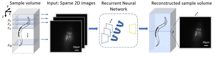
By devising a new recurrent neural network, UCLA researchers have demonstrated a deep learning-enabled volumetric microscopy framework for 3D imaging of fluorescent samples. This new method only requires a few 2D images of the sample to be acquired for reconstructing its 3D image, providing ~30-fold reduction in the number of scans required to image a fluorescent volume. The convolutional recurrent neural network that is at the heart of this 3D fluorescence imaging method intuitively mimics the human brain in processing information and storing memories, by consolidating frequently appearing and important object information and features, while forgetting or ignoring some of the redundant information. Using this recurrent neural network scheme, UCLA researchers successfully incorporated spatial features from multiple 2D images of a sample to rapidly reconstruct its 3D fluorescence image.

Published in Light: Science and Applications, the UCLA team demonstrated the success of this volumetric imaging framework using fluorescent C. Elegans samples, which are widely used as a model organism in biology and bioengineering. Compared with standard wide-field volumetric imaging that involves densely scanning of samples, this recurrent neural network-based image reconstruction approach provides a significant reduction in the number of required image scans, which also lowers the total light exposure on the sample. These advances offer much higher imaging speed for observing 3D specimen, while also mitigating photo-bleaching and phototoxicity related challenges that are frequently observed in 3D fluorescence imaging experiments of live samples.

###

This research is led by Professor Aydogan Ozcan, an associate director of the UCLA California NanoSystems Institute (CNSI) and the Volgenau Chair for Engineering Innovation at the electrical and computer engineering department at UCLA. The other authors include graduate students Luzhe Huang, Hanlong Chen, Yilin Luo and Professor Yair Rivenson, all from electrical and computer engineering department at UCLA. Ozcan also has UCLA faculty appointments in bioengineering and surgery, and is an HHMI professor.

Media Contact
Aydogan Ozcan
[email protected]

Related Journal Article

http://dx.doi.org/10.1038/s41377-021-00506-9

Tags: Chemistry/Physics/Materials SciencesOptics
Share13Tweet8Share2ShareShareShare2

Related Posts

Dual-Frequency Photoacoustic CT Reveals Clear Whole Brain, Centimeters Deep

Dual-Frequency Photoacoustic CT Reveals Clear Whole Brain, Centimeters Deep

July 14, 2026
World-First Neutron Lens Sharply Enhances Atomic-Scale Observations

World-First Neutron Lens Sharply Enhances Atomic-Scale Observations

July 14, 2026

Unveiling the Metallic Secrets of an Ancient Bio-Metal Maw

July 14, 2026

Programmable Arene Ring Opening Enables Diverse Phenol Synthesis

July 14, 2026
Please login to join discussion

POPULAR NEWS

  • New Drug Candidate Developed at McMaster Shows Potential for Treating Brain Cancer

    58 shares
    Share 23 Tweet 15
  • Detection of EDCs in Breast Milk and Infant Urine Up to Six Months Highlights Early Exposure Risks

    77 shares
    Share 31 Tweet 19
  • Experimental Therapy Simultaneously Destroys Prostate Tumor Cells and Reactivates Antitumor Immunity

    46 shares
    Share 18 Tweet 12
  • 高齢者の骨粗鬆症治療の持続性比較

    51 shares
    Share 20 Tweet 13

About

We bring you the latest biotechnology news from best research centers and universities around the world. Check our website.

Follow us

Recent News

Uncanny Valley Effect Detected in Macaques Using 3D Animated Monkey Avatars

Dual-Frequency Photoacoustic CT Reveals Clear Whole Brain, Centimeters Deep

Researchers Identify Therapeutic Vulnerability in Aggressive Childhood Leukemia

Subscribe to Blog via Email

Enter your email address to subscribe to this blog and receive notifications of new posts by email.

Join 85 other subscribers
  • Contact Us

Bioengineer.org © Copyright 2023 All Rights Reserved.

Welcome Back!

Login to your account below

Forgotten Password?

Retrieve your password

Please enter your username or email address to reset your password.

Log In
No Result
View All Result
  • Homepages
    • Home Page 1
    • Home Page 2
  • News
  • National
  • Business
  • Health
  • Lifestyle
  • Science

Bioengineer.org © Copyright 2023 All Rights Reserved.