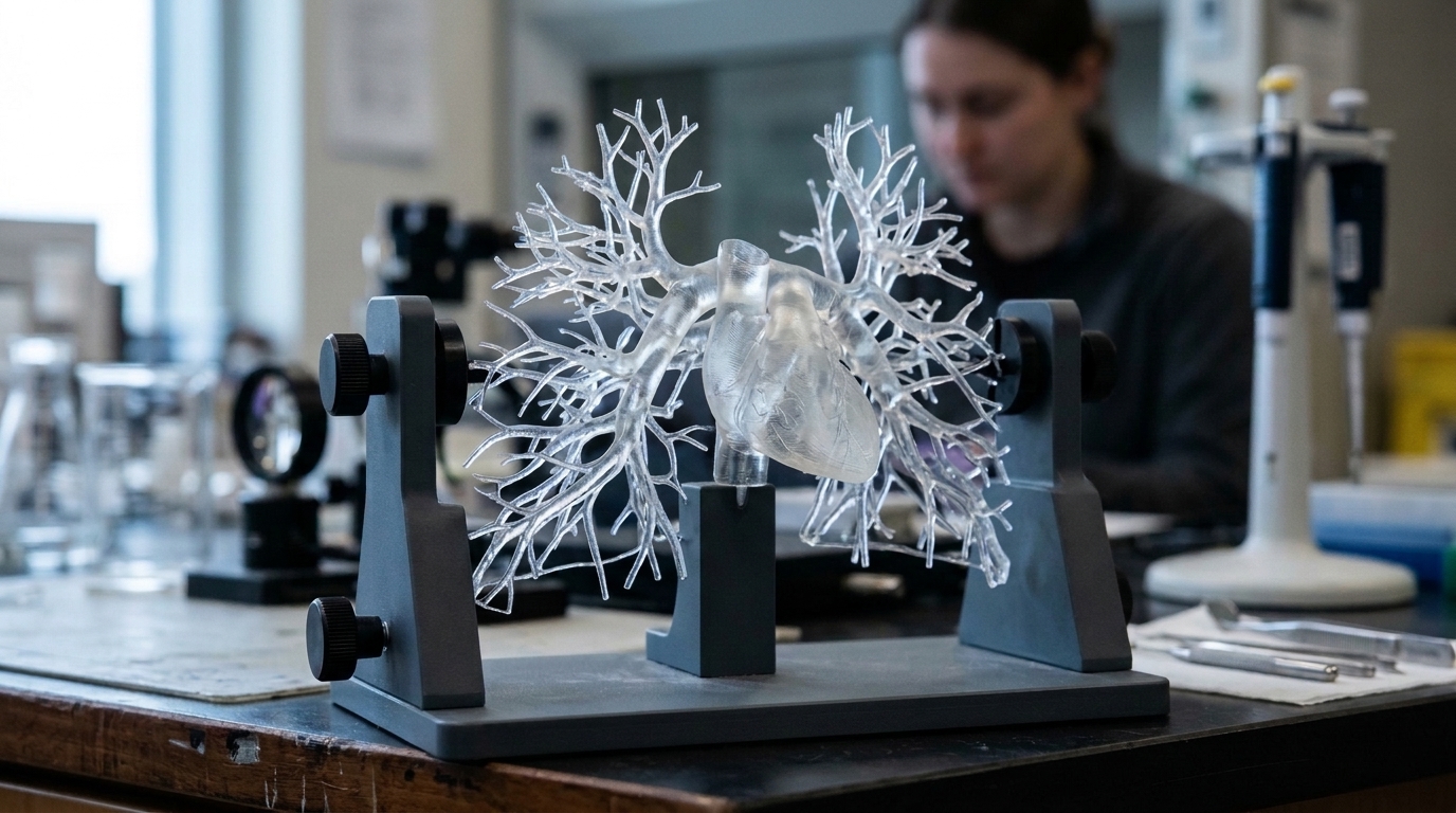
A customizable 3D-printed lung phantom reliably replicates pulmonary embolism conditions for cross-modality diagnostic imaging evaluations, according to research published in 3D Printing in Medicine. The primary objective of the investigation was to produce an anatomically realistic lung phantom containing a simulated pulmonary embolism region, specifically tailored to support the inter-modality study of the pulmonary vascular obstruction index.
In clinical practice, establishing an accurate diagnosis of pulmonary embolism requires specialized medical imaging, with several distinct diagnostic modalities routinely deployed to evaluate patients. These clinical approaches include nuclear medicine techniques, such as planar scintigraphy and positron emission tomography, along with computed tomography techniques, specifically iodine mapping. Creating a reliable experimental method to compare these diverse modalities in assessing functional impairment is vital, as it enables more precise determination of the pulmonary vascular obstruction index. This measurement is clinically critical because the obstruction index has emerged as an independent predictor of disease recurrence in patients diagnosed with unprovoked pulmonary embolism.
To establish a consistent platform for cross-modality comparison, the researchers engineered a dedicated pulmonary embolism lung model incorporating a 5% pulmonary vascular obstruction index. The anatomical geometry of the model was established using a computed tomography scan of a healthy lung phantom alongside human lung segmentation techniques. The physical prototype was manufactured through selective laser sintering using Nylon 12 powder. The investigators specifically selected this powder material and additive manufacturing technique to guarantee complete watertightness across the finished phantom structure during experimental imaging procedures.
The research team validated the physical prototype across five critical evaluation parameters: geometric consistency, density assessment, multi-modality imaging evaluation, motion management, and production cost analysis. When tested across planar scintigraphy, positron emission tomography, and computed tomography iodine mapping, the simulated diagnostic images produced by the model were determined to be highly realistic across all evaluated modalities. Furthermore, the accompanying financial assessment confirmed that the total production cost of the physical model remained under one thousand euros, demonstrating an affordable fabrication workflow.
By delivering highly realistic simulated images across planar scintigraphy, positron emission tomography, and iodine mapping, the model establishes a dependable testing framework for medical imaging technologies.
Subject of Research: 3D-printed lung phantom for pulmonary embolism imaging
Article Title: Pulmonary embolism 3D printing model for inter-modality imaging study
Article References: Tripet, P., Pavoine, M., Le Roux, P.-Y., Guigo, S., Dissaux, B., & Bourhis, D. (2026). Pulmonary embolism 3D printing model for inter-modality imaging study. 3D Printing in Medicine. https://doi.org/10.1186/s41205-026-00354-6
Image Credits: AI Generated
DOI: 10.1186/s41205-026-00354-6
Keywords: pulmonary embolism, 3D printing, lung phantom, computed tomography, nuclear medicine, planar scintigraphy, positron emission tomography, iodine mapping, Pulmonary, embolism, printing, model
News Source: Denise Maddox. (November 5, 2026). Realistic 3D-Printed Lung Model Evaluates Pulmonary Embolism Across Imaging Modalities. Scienmag.