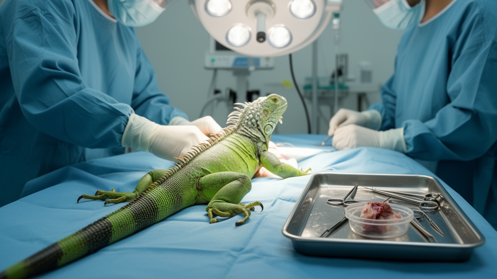
A fourteen-year-old, 3.9-kilogram female green iguana (Iguana iguana) kept under human care became the subject of a rare veterinary diagnosis after arriving at a clinic in Germany with eight weeks of persistent apathy, anorexia and progressively worsening bilateral coelomic distension. Physical examination revealed poor body condition and marked deterioration of her general state. The case, published in Frontiers in Veterinary Science, describes a neoplastic disease that has rarely been reported in iguanas and argues that ovarian tumors deserve a place among the leading differential diagnoses for reproductive disorders in female lizards.
The diagnostic workup began with blood chemistry, radiography and ultrasonography. Blood samples drawn from the ventral tail vein under manual restraint showed glutamate dehydrogenase activity that appeared mildly increased at 12.1 U/L, though the authors caution that validated species-specific reference intervals for this liver enzyme in green iguanas are lacking, making the finding difficult to interpret. Mild hyperuricemia was also detected, with uric acid at 7.81 mg/dl against a reference range of 0.9 to 6.7 mg/dl. Dorsoventral and horizontal beam lateral radiographs revealed marked soft tissue opacity in the median and caudal coelom with a concurrent loss of normal coelomic detail, a pattern consistent with a large space-occupying process.
Ultrasonography, performed with the iguana in dorsal recumbency using a 5 to 9 MHz micro curved array transducer, added crucial detail. Scans from a ventral coupling site showed a large volume of free intracoelomic fluid containing multifocal, moderate corpuscular elements, together with multiple hyperechogenic, heterogeneous and partially vascularized masses in the caudal coelom. Based on these images, neoplasia became the primary differential diagnosis, although the organ of origin could not be clearly identified.
To characterize the fluid further, veterinarians performed an ultrasound-guided coelomic centesis through a caudolateral site on the left coelom. They aspirated 200 milliliters of brownish, serous fluid, and repeated the procedure once daily for three consecutive days, removing 200 milliliters each time. Cytological examination of stained aliquots revealed cocci, heterophilic granulocytes and occasional degranulated mast cells, findings considered consistent with an inflammatory process within the coelomic cavity. Fluid analysis showed a total protein concentration of 3.55 g/dl, albumin of 1.1 g/dl and urea of 7 mg/dl. The authors note that the detection of urea may indicate contamination with urinary material, but the comparatively low urea concentration did not allow a definitive conclusion about the fluid’s origin.
The iguana was hospitalized for three days of preoperative stabilization. Supportive therapy included intravenous fluids at 15 mL/kg once daily, the analgesic meloxicam at 0.3 mg/kg subcutaneously, and the antibiotic marbofloxacin at 10 mg/kg subcutaneously. Hepatic and renal support was provided with phytotherapeutic medications, and the animal was maintained within her preferred optimal temperature zone. Despite this care, no clinical improvement was observed, and the team proceeded to an exploratory coeliotomy, the surgical opening of the coelomic cavity to identify and, if possible, remove the pathology.
General anesthesia was induced with a combination of hydromorphone, alfaxalone, ketamine and medetomidine, then maintained with 1 to 2 percent isoflurane in oxygen delivered through intermittent positive pressure ventilation after intubation with a modified 9.5 feeding tube. Monitoring included Doppler heart rate assessment and continuous cloacal temperature measurement, with heating mats and supplemental pads maintaining body warmth. Positioned in lateral recumbency and aseptically prepared with iodine and chlorhexidine, the iguana received a transverse skin incision on the left flank. Upon entering the coelomic cavity, surgeons aspirated approximately 400 milliliters of brownish, serous ascites fluid. Because the mass was difficult to access, an additional cranial relieving incision was made to improve exposure and permit complete resection.
The ovarian mass itself measured 5 by 4.5 by 4.5 centimeters and was solid, firm and nodular. Its vascular supply was ligated with bipolar electrocautery and the tumor was completely excised. Multiple irregular, ovoid, firm, whitish masses of roughly 3 by 2 by 2.5 centimeters were distributed multifocally within the coelomic cavity, several adherent to the mesentery and coelomic membranes. All were dissected free except one inseparably attached to the greater curvature of the stomach. Extensive fibrinous deposits were removed from the coelomic membrane, the cavity was lavaged with 200 milliliters of warm isotonic crystalloid solution, and a thorough inspection found no additional masses before stepwise closure.
The postoperative course was fatal. The iguana did not regain spontaneous respiration, and intermittent positive pressure ventilation was maintained for a total of three hours. Despite continuous heart rate monitoring and supportive care within an appropriate temperature range, cardiac activity ceased and the animal died approximately three hours after surgery. The authors attribute the death most likely to multifactorial circulatory failure with a potentially hypovolemic component stemming from fluid and protein loss. Before surgery, roughly 600 milliliters of coelomic fluid had already been removed from the 3.9-kilogram animal, equivalent to about 154 mL/kg body weight. Given the marked preoperative deterioration and potential hypovolemia, the additional cardiovascular stress of anesthesia may have contributed to circulatory instability, although the exact mechanism could not be determined retrospectively. The authors also state that, given the severely compromised condition at presentation, euthanasia would have been a reasonable alternative to surgery.
Histopathological examination of the excised tissue clarified the diagnosis. The sample contained remaining ovarian tissue heavily infiltrated with macrophages, epithelioid cells and fewer lymphocytes, often arranged around eosinophilic proteinaceous material most likely of follicular origin, alongside a multinodular mass. That mass consisted of different tissue types organized in a haphazard pattern: nests and islands of partially keratinizing epithelium forming keratin-filled cysts, immature and mature cartilage, adipose tissue and fibrous connective tissue. The tissues showed mild anisocytosis and anisokaryosis with a low mitotic count of fewer than one mitotic figure per 2.37 mm². Together these features established an ovarian teratoma and severe, chronic, multifocal lipogranulomatous oophoritis. Teratomas are tumors composed of tissues derived from two or more germ cell layers, including ectoderm, mesoderm and endoderm, and can contain cartilage, nervous tissue, adipose tissue, hair follicles, respiratory epithelium and glandular tissue. They most commonly arise in the gonads but can occur at extragonadal sites such as the central nervous system, sacrum or mediastinum.
The case sits within a broader context of rising recognition of reptile cancer. Neoplastic disease in reptiles was once considered less common than in mammals, but reported prevalence varies widely with study populations and data sources. A large-scale retrospective study found a prevalence of 2.63 percent among all lizards presented at a specialized teaching hospital, while another retrospective analysis of diagnostic samples from clinically affected lizards, based on 690 samples, found neoplastic disease in 22.9 percent of cases and documented numerous iguana neoplasms including ovarian adenocarcinoma. Teratomas have been described sporadically in lizards, with one reptilian neoplasia study reporting a teratoma prevalence of 1.2 percent in lizards. Previous iguana cases include both benign and malignant ovarian teratomas, an adenocarcinoma and a papillary cystadenocarcinoma, and teratomas have also been reported in the Fiji island banded iguana, garter snake, red-eared slider turtle and bearded dragon. The authors emphasize that the clinical signs in this case, anorexia, apathy and bilateral coelomic distension, overlap with follicular stasis and other reptile diseases such as renal disease, dystocia or viral infections, making further diagnostic work-up essential. They also note that recurrent dystocia has previously been reported in a green iguana with a teratoma, and that in this case it remains unclear whether absent oviposition was associated with dystocia or whether tumor growth led to follicular stasis. When treatment is pursued, complete surgical excision is considered the primary therapeutic approach for teratomas, and surgical resection has proven effective and life-prolonging in previously published case reports, a reminder of how much hinges on timely diagnosis before a patient’s condition deteriorates beyond the limits of anesthesia.
Subject of Research: Diagnosis and surgical removal of an ovarian teratoma with concurrent lipogranulomatous oophoritis in a pet green iguana
Article Title: Case Report: Diagnosis and surgical procedure of an ovarian teratoma in a green iguana (Iguana iguana)
Article References: Zdzuj, V., Hetterich, J., Puff, C., & Pees, M. (2026). Case Report: Diagnosis and surgical procedure of an ovarian teratoma in a green iguana (Iguana iguana). Frontiers in Veterinary Science, 13, Article 1970094. https://doi.org/10.3389/fvets.2026.1970094
Image Credits: AI Generated
DOI: 10.3389/fvets.2026.1970094
Keywords: green iguana, ovarian teratoma, reptile oncology, coeliotomy, oophoritis, ascites, veterinary surgery, histopathology, follicular stasis, differential diagnosis, Iguana iguana, case report
News Source: Ophelia Keating. (October 29, 2026). Rare Ovarian Teratoma Diagnosed in a Green Iguana After Fatal Surgery. Scienmag.