


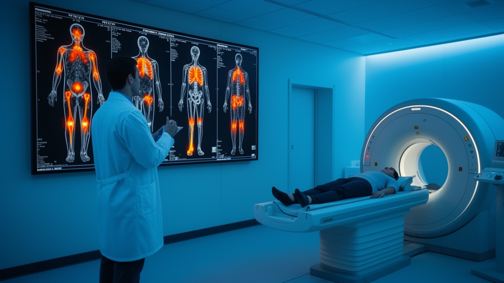
A 46-year-old man scanned for an entirely unrelated cancer mystery has ended up supplying one of medicine’s rarest diagnoses. Together, those two findings define Mazabraud’s syndrome, a benign genetic disorder so uncommon that fewer than 150 cases have been reported in the medical literature to date. The case, published as an open-access report in EJNMMI Reports, illustrates how a scanning technology usually deployed for oncologic staging can stumble upon, and then confidently characterize, a condition that most clinicians will never encounter in an entire career.
The patient’s medical history already read like a diagnostic puzzle. Twenty years earlier he had undergone resection of a myxoma in his right thigh after noticing enlargement of the limb, but he received no regular follow-up afterward and remained symptom-free in the intervening decades. One year before the PET/CT study, a thyroid nodule led to a right lobectomy with central neck dissection for thyroid cancer. Postoperative pathology revealed papillary thyroid carcinoma that had spread to one of two lymph nodes in the right central neck compartment. More troubling still, metastatic squamous cell carcinoma was identified in a separate lymph node in the suprasternal fossa. Subsequent head and neck MRI, nasopharyngoscopy, and laryngoscopy all failed to show any abnormal findings, so the clinical team suspected a metastatic squamous cell carcinoma of unknown primary origin, an entity abbreviated CMSCCUP.
That diagnosis set the stage for the incidental discovery. FDG PET/CT was performed specifically to identify the primary site of the squamous cell carcinoma. The scan found no occult primary tumor anywhere in the body. What it did find, in the right thigh, was a cluster of multiple well-defined, homogeneous, hypodense, round-like nodules sitting in the intermuscular space, with a mean CT value of 17 Hounsfield units and mildly increased, homogeneous FDG uptake, with maximum standardized uptake values ranging from 1.6 to 2.9. Given the patient’s remote history of thigh myxoma resection, these features pointed to recurrence of the intramuscular myxomas rather than anything malignant.
The bone findings completed the syndrome. Fibrous dysplasia was identified in the right femur, tibia, talus, cuneiform bone, and first metatarsal bone. The lesions appeared as multiple well-defined hypodense and ground-glass opacities growing along the medullary cavity, some with sclerotic margins, and focal thinning of the adjacent cortical bone that nonetheless remained intact. Characteristically, the long axis of the lesions ran parallel to that of the long bones. Subsequent percutaneous biopsy of the soft-tissue mass confirmed intramuscular myxoma, and with both components established, a final diagnosis of Mazabraud’s syndrome was made. Detailed cross-sectional images documented the extent of the disease: three round, slightly hypodense masses measuring 1.2 to 3.8 centimeters adjacent to the right pectineus and iliopsoas muscles; an osteolytic lesion with a sclerotic border at the femoral lesser trochanter; and multiple irregular, longitudinal osteosclerotic lesions within the tibial medullary cavity, some extending to the adjacent cortex, with markedly increased FDG uptake at SUVmax values of 3.1 to 8.2. Smaller similar lesions were present in the right talus, cuneiform, and first metatarsal.
Mazabraud’s syndrome is, at its core, a disorder of a single gene. Pathogenetically it arises from inactivating mutations in the GNAS1 gene, which drive abnormal mesenchymal cell proliferation. The syndrome shows a female predominance, a tendency to affect the lower extremities, and a notable right-sided predilection, all of which this male patient’s right-sided, lower-limb disease fits with striking fidelity. Most patients are asymptomatic, although bone deformity or fracture may occur. Intramuscular myxomas are typically multifocal, sit adjacent to severely affected bones, and are mostly located in the lower limbs. Because symptoms are absent or mild, such as slight swelling or bone discomfort, the syndrome is frequently delayed in diagnosis or missed altogether, which is precisely what nearly happened over the two decades between this patient’s original surgery and his scan.
The authors argue that the case demonstrates the classic clinical and imaging features of the syndrome in one body: lower extremity location, multifocal intramuscular myxomas, polyostotic fibrous dysplasia, and right-sided involvement. What makes the report compelling is that the diagnosis was made entirely incidentally during oncologic staging for an unrelated malignancy. Unlike conventional imaging, PET/CT provides integrated whole-body metabolic and anatomical information in a single session. On the scan, the myxomas appeared as well-circumscribed, hypodense masses with mild homogeneous FDG uptake, while the bone lesions showed the ground-glass intramedullary pattern with sclerotic margins and cortical thinning that is characteristic of fibrous dysplasia. Those combined features allowed reliable differentiation from malignant bone lesions, which instead display cortical destruction, soft-tissue masses, and infiltrative growth.
The differential diagnosis carries real clinical weight. A major challenge, the authors note, is distinguishing the intramuscular myxomas of Mazabraud’s syndrome from low-grade soft tissue sarcomas and from soft tissue metastases. Low-grade sarcomas typically present as large, infiltrative masses with ill-defined borders, heterogeneous density, and focal hypermetabolism suggestive of dedifferentiation. Soft tissue metastases usually occur in patients with a known primary malignancy, show multifocal involvement, and demonstrate markedly increased FDG uptake. In this patient, who genuinely did harbor a concurrent malignancy, the mild, homogeneous FDG uptake and the typical CT features of both the myxomas and the fibrous dysplasia effectively excluded malignant processes and prevented misdiagnosis, an outcome the authors describe as particularly critical in patients with concurrent cancers to avoid inappropriate treatment. The report also acknowledges an important caveat: magnetic resonance imaging is regarded as the preferred modality for the accurate diagnosis of Mazabraud’s syndrome, but its limited scanning coverage tends to result in missed lesions at distant anatomical sites, which is exactly where whole-body PET/CT holds an advantage.
The triple coexistence of Mazabraud’s syndrome, papillary thyroid carcinoma, and metastatic squamous cell carcinoma of unknown primary is extraordinarily rare, and the authors are careful not to overinterpret it. Coexistence of Mazabraud’s syndrome with malignant neoplasms, although rare, has been reported in several previous cases, and its specific mechanism requires further investigation. The syndrome is associated with inactivating mutations of the GNAS1 gene, and in some rare cases the mechanism underlying malignant transformation of the syndrome has also been linked to GNAS R201H/C mutations. However, targeted genetic testing for GNAS or other susceptibility loci was not performed in this patient, who declined genetic testing as well as regional targeted therapy. Current evidence, the authors state, is insufficient to confirm a shared genetic susceptibility via the GNAS pathway between this syndrome and non-endocrine malignancies or thyroid carcinoma, and it remains unclear whether the triple co-occurrence represents a sporadic event. They suggest that the potential role of the GNAS pathway in multineoplastic processes warrants further investigation.
The oncologic workup itself ended inconclusively but reassuringly in the short term. For the CMSCCUP, FDG PET/CT did not detect an occult primary tumor. The patient entered routine oncologic follow-up, and during short-term surveillance no distant metastasis and no primary tumor were identified.
The report’s conclusion is measured. Mazabraud’s syndrome is a rare benign entity that is easily overlooked because of its non-specific manifestations, and FDG PET/CT enables whole-body metabolic and anatomical evaluation that can simultaneously detect intramuscular myxomas and fibrous dysplasia, the hallmark combination of the syndrome, while facilitating differentiation from malignant lesions and thereby preventing misdiagnosis and inappropriate clinical decisions. For patients found to have the syndrome alongside concurrent malignancies, the authors recommend long-term multidisciplinary surveillance. In an era when PET/CT is most often discussed as a staging tool for cancer, the case is a reminder that the same whole-body view can surface benign genetic disease hiding in plain sight for twenty years, provided the interpreting physician recognizes a pattern that fewer than 150 reported cases have taught medicine to see.
Subject of Research: Incidental diagnosis of Mazabraud's syndrome by FDG PET/CT during oncologic staging for metastatic squamous cell carcinoma of unknown primary
Article Title: A case of Mazabraud’s syndrome identified by FDG PET/CT
Article References: Liu, J., Li, X., Fang, J., Cai, H., & Yi, H. (2026). A case of Mazabraud’s syndrome identified by FDG PET/CT. EJNMMI Reports, 10(1), Article 34. https://doi.org/10.1186/s41824-026-00312-9
Image Credits: AI Generated
DOI: 10.1186/s41824-026-00312-9
Keywords: Mazabraud's syndrome, FDG PET/CT, intramuscular myxoma, fibrous dysplasia, GNAS1 mutation, case report, cancer of unknown primary, papillary thyroid carcinoma, soft tissue sarcoma differential, whole-body imaging, benign genetic disorder, incidental finding
News Source: Nathaniel Bowman. (October 29, 2026). Rare Mazabraud’s Syndrome Uncovered Incidentally on FDG PET/CT During Cancer Staging. Scienmag.



