Sebaceous adenoma of the eyelid represents an exceptionally rare entity within dermatopathology, characterized by its benign yet diagnostically nuanced nature. This uncommon tumor arises from the sebaceous glands, which are specialized structures responsible for lipid secretion within the skin. Despite the prevalence of sebaceous glands throughout the integumentary system, sebaceous adenomas comprise less than 0.5% of all cutaneous neoplasms and account for a small fraction—approximately 1 to 2%—of eyelid tumors, highlighting their rarity in ocular adnexal pathology.
The recent case report published in Oncoscience offers an in-depth clinical, histopathological, and immunohistochemical examination of sebaceous adenoma localized specifically to the eyelid, demonstrating the complexities involved in distinguishing this benign tumor from both benign papillomatous lesions and more sinister malignant counterparts such as sebaceous carcinoma. The case underscores the importance of a multidisciplinary diagnostic approach integrating clinical observations with microscopic and molecular evaluations to avoid misdiagnosis.
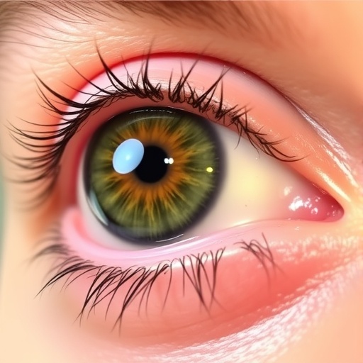
In clinical presentation, sebaceous adenoma typically manifests as a slow-growing, yellowish exophytic mass with a papillomatous surface on the lower eyelid. The lesion’s gradual enlargement over years and its distinct coloration are suggestive but not pathognomonic. This clinical ambiguity necessitates histopathological analysis, as visual inspection alone can be misleading. The papillomatous morphology complicates clinical diagnosis, as it mimics other benign elevations and can be easily confounded with malignant tumors causing significant concern for practitioners.
Histologically, sebaceous adenoma is typified by sharply demarcated lobules composed predominantly of mature sebocytes displaying characteristic vacuolated cytoplasm, indicative of lipid-rich cellular content. Surrounding these sebocytes is a peripheral rim of basaloid cells, which are smaller, darker-staining cells with a high nuclear-to-cytoplasmic ratio, reflective of proliferative precursors in sebaceous differentiation. Crucially, the absence of nuclear atypia, necrosis, and mitotic figures within these samples corroborates the lesion’s benign nature and aids in differentiating it from sebaceous carcinoma, which would exhibit cytologic atypia and invasive growth patterns.
Immunohistochemical profiling emerges as a vital adjunct to traditional histology in delineating sebaceous adenoma. The tumor cells demonstrate positivity for epithelial membrane antigen (EMA), a glycoprotein expressed on the membrane of differentiated epithelial cells, including sebocytes, confirming their sebaceous origin. Concurrent low Ki-67 labeling index—under 5% in this case—signifies minimal proliferative activity characteristic of benign lesions and contrasts starkly with malignant sebaceous tumors that usually display elevated Ki-67 indices reflecting uncontrolled cell division.
Differential diagnosis of eyelid tumors mandates careful evaluation because treatment strategy and prognosis vary markedly between benign and malignant entities. Sebaceous adenomas, while benign, necessitate excision with clear margins to prevent potential recurrence but do not require aggressive interventions such as wide local excision or adjuvant therapy reserved for carcinoma. Misinterpretation could lead to overtreatment or conversely, inadequate management if malignancy is overlooked.
Beyond immediate diagnostic concerns, the case also raises the significant consideration of hereditary cancer syndromes, particularly Muir–Torre syndrome (MTS). MTS is characterized by sebaceous neoplasms associated with internal malignancies due to defects in DNA mismatch repair genes, including MLH1 and MSH2. Although this patient lacked a personal or family history indicative of MTS, awareness and appropriate screening become imperative when sebaceous neoplasms present in younger individuals or in multiple anatomical locations to exclude syndromic associations.
This investigation enriches existing medical literature by presenting a comprehensive examination of sebaceous adenoma’s unusual papillomatous variant localized to the eyelid. It emphasizes the crucial synergy between clinical assessment, detailed histopathology, and sophisticated immunohistochemistry for accurate diagnosis. Furthermore, it highlights the need for heightened diagnostic vigilance to distinguish benign from malignant sebaceous lesions, thereby optimizing patient management.
The report delineates the clinical trajectory of an elderly patient exhibiting a lesion whose initial suspicion leaned toward other benign or potentially malignant eyelid pathologies. The delayed growth pattern and clinical features typify sebaceous adenoma yet also stress the importance of contemplating a broad differential diagnosis in dermatological oncology, particularly in atypical presentations. This nuanced diagnostic pathway prevents under- or over-treatment and guides the formulation of personalized care strategies.
Histopathological details illuminate the microarchitecture of this adenoma, echoing classical sebaceous biology, while the immunohistochemical markers employed provide molecular confirmation of cellular differentiation. EMA positivity confirms glandular epithelial lineage, whereas the low Ki-67 index corroborates the benign proliferative status. Such immunoprofiles are indispensable tools in contemporary pathology, particularly in cases where histology alone may provide equivocal results.
Significantly, this case exemplifies the subtlety required in recognizing dermatologic tumors on the eyelid—a region with complex anatomy and a diverse spectrum of neoplastic processes. Eyelid tumors encompass a wide array of benign, premalignant, and malignant lesions, each demanding distinct therapeutic pathways. Therefore, detailed pathological and immunohistochemical studies are critical in distinguishing entities like sebaceous adenoma from sebaceous carcinoma, basal cell carcinoma, and squamous cell carcinoma, which pose greater morbidity.
In summary, the case report not only contributes to clinical awareness of sebaceous adenoma’s clinical and pathological spectrum but also serves as a reminder of the essential role of integrated diagnostics in dermatology and oncology. The uncommon occurrence of sebaceous adenomas on the eyelid, coupled with their potential to mimic malignant lesions, necessitates meticulous clinical scrutiny supported by advanced laboratory analyses and an understanding of associated hereditary syndromes for comprehensive patient care.
Subject of Research: People
Article Title: Sebaceous adenoma of the eyelid: A clinical, histopathological, and immunohistochemical perspective
News Publication Date: April 2, 2026
Web References: https://doi.org/10.18632/oncoscience.655
Image Credits: Copyright: © 2026 Rathod et al. Licensed under Creative Commons Attribution License (CC BY 4.0)
Keywords: sebaceous adenoma, eyelid tumor, histopathology, immunohistochemistry, epithelial membrane antigen, Ki-67, sebaceous glands, benign neoplasm, sebaceous carcinoma differential, Muir–Torre syndrome, cutaneous neoplasms, ocular oncology
Tags: benign eyelid neoplasmsclinical features of eyelid tumorsdermatopathology of sebaceous glandsdifferential diagnosis sebaceous carcinomahistopathological assessment of eyelid lesionsimmunohistochemical analysis in dermatologymultidisciplinary tumor diagnosisocular adnexal pathologypapillomatous eyelid lesionsrare eyelid tumorssebaceous adenoma of eyelidsebaceous gland tumors diagnosis

