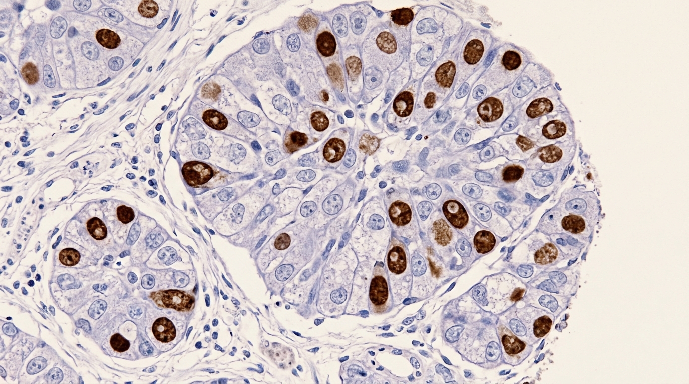
In a comprehensive retrospective study examining 10,471 primary breast cancer cases diagnosed between 2021 and 2024 at Nanchang People’s Hospital, researchers identified primary acinic cell carcinoma of the breast as an exceptionally rare triple-negative malignancy with an institutional incidence of 0.085 percent, defined by distinctive microglandular and acinar architectures and a consistent mutant-type p53 protein expression pattern. Classified within the fifth edition World Health Organization guidelines as a salivary gland-type breast tumor, this uncommon neoplasm accounts for fewer than one-tenth of one percent of breast malignancies, with approximately seventy cases previously reported in global literature. The investigation evaluated surgical resection specimens from nine confirmed patients to clarify the clinical presentation, histomorphological spectrum, immunohistochemical profile, and therapeutic outcomes of this elusive tumor type.
The research team initiated case selection through the institutional pathology database, searching for entries matching breast acinic cell carcinoma and salivary gland-type tumors, which retrieved eleven suspected cases across the four-year study window. Following the application of strict inclusion and exclusion criteria, two candidate cases were excluded: one due to an uncertain pathological diagnosis and another owing to concurrent invasive ductal carcinoma, yielding a final cohort of nine primary breast acinic cell carcinoma patients. Diagnostic confirmation required independent evaluation by two senior pathologists with at least ten years of clinical experience, with complete diagnostic consensus. Enrolled cases required complete demographic, imaging, surgical, and therapeutic records with a follow-up of at least three months, while secondary metastases from salivary gland sites were excluded.
All nine patients in the study cohort were female, ranging in age from 37 to 58 years with a median age of 47 years. Primary tumors occurred in the right breast in five cases and the left breast in four cases. Anatomical distribution showed that eight lesions were situated within the upper outer quadrant, with only one lesion located in the lower outer quadrant. Clinically, six tumors presented as hard masses with poorly demarcated borders and restricted mobility, whereas three presented as firm masses with well-circumscribed borders and good mobility. Tumor diameter ranged from 1.5 to 4.5 centimeters, averaging 2.82 centimeters. Preoperative ultrasound assessments classified the lesions as BI-RADS 4A in four cases, 4B in three, 4C in one, and 6 in one.
Gross examination of surgical resection specimens revealed solitary masses measuring between 1.5 and 4.5 centimeters, displaying firm to tough textures, ill-defined margins, and cut surfaces colored gray-white, gray-yellow, or gray-red, with no well-formed capsule observed in any case. Histopathologically, the series comprised five cases of pure acinic cell carcinoma and four cases of mixed acinic cell carcinoma. Pure cases were defined as tumors composed exclusively or predominantly of classic morphology displaying microglandular or acinar patterns with diastase-resistant periodic acid-Schiff-positive cytoplasmic granules, triple-negative receptor status, and corroborative positive staining for amylase, lysozyme, and alpha-1-antichymotrypsin. Mixed cases were defined as tumors where classic acinic components comprised between ten and ninety percent of total tumor volume coexisting with another distinct invasive breast carcinoma subtype.
Microscopic architectural analysis of the acinic carcinoma components demonstrated invasive growth with diverse structural subtypes across the cohort. Predominant growth patterns were microglandular and acinar, accompanied by less frequent microcystic, follicular, and solid architectures, with eosinophilic secretions universally present within glandular lumens. Cytologically, tumor cells displayed round, oval, or polygonal contours with moderate to abundant cytoplasm showing granular, basophilic, or clear appearances. Nuclei were round, oval, or irregular with finely granular, coarsely granular, or vesicular chromatin, prominent nucleoli, and identifiable mitotic figures. In the four mixed cases, coexisting non-acinic components comprised high-grade triple-negative carcinomas, including two cases of invasive ductal carcinoma of no special type, one case of invasive papillary carcinoma, and one case of matrix-producing metaplastic carcinoma.
Standardized immunohistochemical staining was conducted using the streptavidin-biotin-peroxidase method on four-micrometer formalin-fixed, paraffin-embedded tissue sections according to manufacturer protocols. Following xylene deparaffinization and endogenous peroxidase quenching with hydrogen peroxide, heat-mediated antigen retrieval was performed prior to overnight primary antibody incubation at four degrees Celsius. Slides were treated with secondary antibody detection reagents for thirty minutes at room temperature, followed by diaminobenzidine visualization and hematoxylin counterstaining before mounting with neutral balsam. Phosphate-buffered saline replaced primary antibodies in negative controls, while established positive breast cancer tissues served as positive controls. The primary antibody panel evaluated estrogen receptor, progesterone receptor, androgen receptor, Ki-67, p53, human epidermal growth factor receptor 2, lysozyme, alpha-1-antichymotrypsin, collagen IV, epithelial membrane antigen, and S100 protein across all cases.
Interpretation of immunohistochemical results followed established international consensus criteria. Nuclear staining in under one percent of tumor cells defined negative estrogen and progesterone receptor expression under American Society of Clinical Oncology and College of American Pathologists guidelines, while androgen receptor positivity required nuclear staining in at least ten percent of cells. Human epidermal growth factor receptor 2 evaluation classified absent or faint membrane staining in ten percent or fewer cells as score zero, faint incomplete staining in over ten percent as score one-plus, and equivocal staining as score two-plus. Proliferative activity was categorized using a twenty percent Ki-67 threshold, and p53 status was assessed as wild-type or mutant-type via PROMISE criteria. Collagen IV staining evaluated basement membrane continuity, while lysozyme, alpha-1-antichymotrypsin, and epithelial membrane antigen were scored semi-quantitatively using an H-score cut-off of 150.
Immunohistochemical profiling demonstrated a characteristic biomarker profile across all nine cases. All tumors showed complete negativity for estrogen receptor and androgen receptor, while progesterone receptor was weakly positive in two cases and negative in seven. Human epidermal growth factor receptor 2 staining was negative in five cases and scored one-plus in four cases, with no equivocal two-plus cases requiring fluorescence in situ hybridization. Lysozyme was positive in eight cases and negative in one, while alpha-1-antichymotrypsin was positive in four cases and negative in five. Epithelial membrane antigen demonstrated diffuse, strong positivity in all cases, and S100 protein was positive in seven cases. Notably, both S100-negative cases were positive for lysozyme and alpha-1-antichymotrypsin. All nine cases displayed a mutant-type p53 protein pattern, with Ki-67 proliferation indices ranging from five to eighty percent.
Collagen IV immunohistochemical staining yielded negative results across all nine tumors, confirming the absence of a continuous basement membrane encircling the neoplastic cell nests. This absence provides a critical diagnostic criterion for distinguishing primary acinic cell carcinoma from morphological mimics, particularly microglandular adenosis and atypical microglandular adenosis. While microglandular adenosis and atypical microglandular adenosis share microglandular patterns, triple-negative receptor status, and frequent TP53 mutations with acinic cell carcinoma, they characteristically retain an intact, continuous basement membrane demonstrable by positive collagen IV and laminin immunoreactivity. The shared mutant-type p53 staining observed across microglandular adenosis, atypical microglandular adenosis, and acinic cell carcinoma supports a proposed multi-step oncogenetic pathway, in which breast acinic cell carcinoma represents the invasive malignant endpoint of neoplastic progression arising from low-grade triple-negative glandular precursors.
The study highlighted molecular distinctions separating breast acinic cell carcinoma from its salivary gland counterpart and other salivary gland-like breast tumors. While histologically resembling salivary gland acinic cell carcinoma, the salivary entity is characteristically defined by recurrent NR4A3 alterations, whereas mammary acinic cell carcinoma consistently exhibits mutant-type p53 expression reflecting underlying TP53 gene alterations. Among other rare triple-negative salivary gland-type breast tumors—which include adenoid cystic carcinoma, secretory breast carcinoma, mucoepidermoid carcinoma, and pleomorphic adenoma, collectively accounting for five to ten percent of triple-negative breast cancers—accurate differentiation is essential. Secretory breast carcinoma is typically defined by ETV6-NTRK3 gene fusions and adenoid cystic carcinoma by MYB-NFIB fusions, and neither tumor harbors the recurrent mutant-type p53 expression pattern that characteristically marks primary breast acinic cell carcinoma.
All nine patients underwent surgical intervention as their primary therapeutic modality, with five patients receiving breast-conserving surgery and four undergoing simple mastectomy. Ipsilateral sentinel lymph node biopsy was performed in every case to assess regional nodal involvement, and histological examination revealed no sentinel lymph node metastasis in any patient. Following surgical resection, pathological tumor staging in accordance with the American Joint Committee on Cancer staging system categorized one patient with stage I disease and eight patients with stage II disease.
Regular clinical follow-up for the nine patients ranged from 4 to 48 months post-surgery. Over this monitoring period, no adverse clinical events occurred, with zero instances of local tumor recurrence, distant metastasis, or cancer-related mortality, indicating favorable short-to-medium-term outcomes for this series. Nevertheless, the study authors emphasized that these results should be interpreted cautiously due to the small single-center cohort and relatively limited follow-up duration. Given that the four mixed acinic cell carcinoma cases contained high-grade components—including invasive ductal carcinoma, invasive papillary carcinoma, and matrix-producing metaplastic carcinoma—and considering documented literature reports of distant metastasis and high-grade transformation, extended clinical surveillance remains essential for all patients diagnosed with this rare malignant entity.
Subject of Research: Clinicopathological and immunohistochemical features of primary breast acinic cell carcinoma
Article Title: Primary acinic cell carcinoma of the breast: insights from a nine-case series and comprehensive literature review
Article References: Zhou, G., Yang, L., Gui, X., Qu, W., Huang, A., & Gui, S. (2026). Primary acinic cell carcinoma of the breast: insights from a nine-case series and comprehensive literature review. Diagnostic Pathology, 21(1), Article 65. https://doi.org/10.1186/s13000-026-01832-5
Image Credits: AI Generated
DOI: 10.1186/s13000-026-01832-5
Keywords: acinic cell carcinoma, breast cancer pathology, triple-negative breast cancer, p53 mutation, collagen IV, microglandular adenosis, salivary gland-type breast tumors, lysozyme, alpha-1-antichymotrypsin, epithelial membrane antigen, S100 protein, Ki-67 proliferation index, BI-RADS 4, sentinel lymph node biopsy, breast-conserving surgery, adjuvant chemotherapy, PROMISE molecular typing, Nanchang People's Hospital
News Source: Nathaniel Bowman. (November 11, 2026). Rare Breast Acinic Cell Carcinomas Consistently Exhibit Mutant p53 Expression Patterns. Scienmag.