


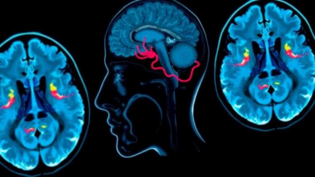
A 47-year-old woman who arrived at an emergency department obtunded, seized continuously, and was later found to have a basilar artery thrombosis has become the subject of a rare documented case in which a brain perfusion scan showed complete absence of blood flow to the posterior fossa while perfusion to both cerebral hemispheres remained intact. The case report, published as open access research in EJNMMI Reports on 14 August 2026, describes how this dissociation between infratentorial and supratentorial circulation initially prevented a declaration of brain death, and how the patient nonetheless progressed to diffuse cerebral edema, massive herniation, and clinical brain death within a day. The authors, Ghazal Shadmani and Amin Haghighat Jahromi, present the clinical course, imaging findings, and outcome to illustrate a presentation so uncommon that only a handful of comparable cases have been reported in the medical literature.
The diagnosis of brain death is primarily clinical and is usually straightforward when both the supratentorial structures of the cerebral hemispheres and the infratentorial structures of the brainstem and cerebellum are irreversibly injured together. The fundamental concept involves a comatose state, loss of all cranial nerve function, and the irreversible loss of spontaneous respiration. Before the diagnosis is made, clinicians must identify the underlying disorder responsible for the brain injury and rule out confounders and reversible causes of coma and apnea, including drug intoxication, hypothermia, acute metabolic disturbances, and neuromuscular disorders. When all criteria are met in the absence of confounders, the patient is considered legally deceased. Isolated catastrophic posterior fossa lesions, particularly those involving the brainstem, are uncommon and may instead demonstrate preserved supratentorial perfusion or cortical electrical activity on ancillary testing even in the absence of any brainstem function.
Two anatomical approaches to determining death by neurologic criteria are recognized globally: whole-brain death and brainstem death. Whole-brain death is typically applied in scenarios involving large supratentorial lesions, such as ischemia, trauma, or hemorrhage, causing progressive transtentorial herniation with eventual brainstem dysfunction, or diffuse brain injury such as post-cardiac arrest hypoxia affecting both supratentorial and infratentorial structures. A distinct clinical situation arises in patients with isolated fatal infratentorial lesions, particularly affecting the brainstem. In these cases, patients may lose all brainstem reflexes while retaining functional supratentorial structures, and ancillary studies including electroencephalography, visual evoked potentials, or brain perfusion scintigraphy may demonstrate preserved cortical activity or supratentorial perfusion even when clinical criteria for brain death are fulfilled. The debate continues internationally as to whether the definition of death by neurologic criteria should require death of the entire brain or only the brainstem.
The rarity of this dissociation is underscored by the figures the authors cite. In a series of 161 brain death cases, Varelas and colleagues reported only three with isolated posterior fossa injury, a prevalence of 1.9 percent. Such lesions can result from basilar artery thrombosis, subarachnoid hemorrhage, massive trauma, or complications of posterior fossa surgery. Although the concept of brain death is relatively consistent internationally, the criteria and the use of ancillary testing vary across countries. In the United Kingdom and some U.S. states, a combination of coma, confirmed apnea, and absent brainstem reflexes is sufficient to declare brain death regardless of lesion location. However, reports of patients with isolated infratentorial injury who retain supratentorial function have prompted consideration of guideline-based ancillary testing to confirm the diagnosis, and the 2023 international consensus guidelines emphasize careful evaluation in primary infratentorial injury and recommend ancillary testing when portions of the clinical examination cannot fully confirm brain death.
The patient in this case was transported to the emergency department in an obtunded state following the sudden onset of right facial droop and aphasia. Her history was notable for one week of migraines and recent heavy pipe tobacco use, and she had no prior history of chronic seizures. Upon arrival she experienced continuous seizure activity and her clinical status rapidly decompensated, requiring emergent intubation. Her National Institutes of Health Stroke Scale score was estimated at 31, indicating devastating neurologic impairment. A non-contrast head CT revealed a dense basilar artery, suggestive of thrombosis, and CT angiography demonstrated distal basilar artery occlusion extending into the right P1 segment. Her family confirmed that the last time she was seen well was approximately three hours prior. She received intravenous tissue plasminogen activator and levetiracetam, and she underwent mechanical thrombectomy, achieving Thrombolysis in Cerebral Infarction grade 2b reperfusion, a partial recanalization, after two passes.
The patient was admitted to the neuro-intensive care unit following the procedure. Because of poor awakening from anesthesia, a follow-up head CT without contrast was obtained, which revealed increased brainstem edema and obstructive hydrocephalus, with decreased size of the fourth ventricle and enlargement of the third and lateral ventricles. Upon neurosurgical evaluation, she was deemed not a candidate for further intervention due to a poor neurological examination and her recent administration of tPA. A subsequent head CT demonstrated evolving edema and infarction involving the bilateral cerebellar hemispheres, the brainstem, and the right occipital lobe, along with widespread cerebral sulci effacement, although gray-white matter differentiation of the frontal, parietal, and anterior temporal lobes was preserved. Her clinical condition continued to deteriorate despite the partially successful endovascular intervention.
The neurologic examination at this point revealed a comatose patient who was unresponsive with eyes closed and no spontaneous eye opening. Her pupils were bilaterally dilated at 6 millimeters and nonreactive. Brainstem reflexes, including corneal responses, horizontal oculocephalic movements, cough, and gag reflexes, were all absent. There were no spontaneous movements of the extremities and no responses to noxious stimulation. The seriousness of her condition and her poor prognosis were discussed with her family members, and the possibility of organ donation was raised, which the family confirmed aligned with the patient’s previously expressed wishes. A brain perfusion study using the lipophilic tracer 99mTc-bicisate was then obtained to evaluate cerebral blood flow at the bedside.
The scintigraphic findings were striking. The anterior cerebral radionuclide angiogram and 20-minute delayed static images demonstrated complete absence of perfusion to the posterior fossa, while cortical perfusion in both cerebral hemispheres was preserved. Brain perfusion scintigraphy is an established adjunctive test for the determination of brain death, particularly in patients in whom clinical examination or EEG is unreliable or cannot be performed, and demonstration of absent intracranial perfusion is highly specific and associated with a grave prognosis, providing a practical alternative to invasive cerebral angiography. In this instance, however, the tracer study showed the reverse of the classic pattern: the infratentorial circulation had vanished while the supratentorial circulation persisted. From a medicolegal standpoint, the authors report, the preserved supratentorial perfusion precluded a declaration of brain death at that time, and life-sustaining treatment was continued.
A follow-up head CT without contrast the next day demonstrated extensive cytotoxic edema involving the brainstem, bilateral cerebellar hemispheres, and occipital lobes, along with signs of upper transtentorial and descending tonsillar herniation and progressive obstructive hydrocephalus. Later that day, the patient was clinically declared brain dead. No additional imaging was performed. Her liver and kidneys were successfully recovered for transplantation. The authors note that preserved supratentorial perfusion in the absence of infratentorial perfusion has been previously described in patients with isolated posterior fossa lesions, and a similar pattern has also been reported in a case of widespread subarachnoid hemorrhage. In previously reported cases, patients were ultimately declared brain dead within a few days, with or without repeated imaging, although the clinical significance of supratentorial perfusion in the setting of severe brainstem damage remains unclear.
The proposed mechanism for the inevitable supratentorial tissue injury is mechanical and hemodynamic: aqueductal obstruction due to progressive brainstem edema leads to hydrocephalus and gradual loss of supratentorial perfusion, while collapse of the vein of Galen due to sustained intracranial pressure may further impair venous outflow, eventually overwhelming the brain’s ability to autoregulate. The authors caution that they did not obtain serial perfusion studies to document the temporal course in this patient, but prior reports suggest that such preservation is often transient and uniformly associated with poor outcomes. The case, they conclude, is a rare example of isolated posterior fossa injury with preserved supratentorial perfusion compatible with brainstem death, and it highlights the importance of ancillary testing in uncommon supratentorial-infratentorial dissociation to differentiate brain death from brainstem death, with implications for accurate and timely determination of death by neurologic criteria and for ethical decision-making around organ donation.
Subject of Research: A case report of isolated brainstem death with preserved supratentorial perfusion on 99mTc-bicisate brain perfusion scintigraphy following basilar artery thrombosis.
Article Title: Isolated brainstem death: case presentation demonstrating preserved supratentorial perfusion on 99mTc-bicisate brain scintigraphy
Article References: Shadmani, G., & Haghighat Jahromi, A. (2026). Isolated brainstem death: case presentation demonstrating preserved supratentorial perfusion on 99mTc-bicisate brain scintigraphy. EJNMMI Reports, 10(1), Article 36. https://doi.org/10.1186/s41824-026-00283-x
Image Credits: AI Generated
DOI: 10.1186/s41824-026-00283-x
Keywords: brain death, brainstem death, basilar artery thrombosis, 99mTc-bicisate, brain perfusion scintigraphy, posterior fossa, mechanical thrombectomy, obstructive hydrocephalus, ancillary testing, organ donation, death by neurologic criteria, case report
News Source: Ophelia Keating. (October 29, 2026). Rare Brainstem Death Case Shows Preserved Supratentorial Perfusion on Brain Scan. Scienmag.



