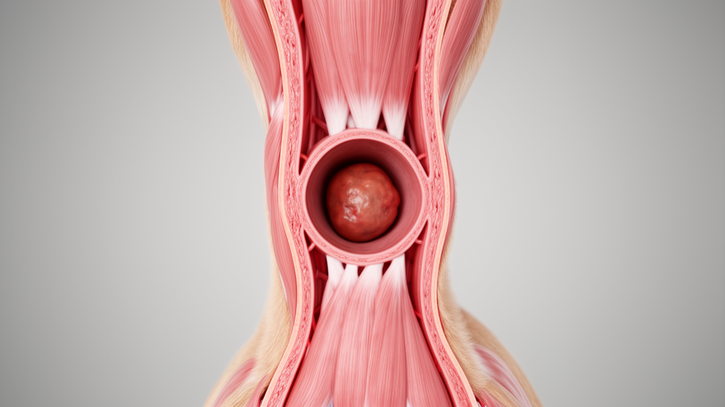
Researchers have documented the first comprehensive pathological and immunohistochemical characterization of a histologically benign chemodectoma arising specifically from the abdominal aorta trifurcation in a dog. The study details the clinical presentation, diagnostic imaging, surgical challenges, and detailed histopathological findings of a 9-year-old spayed female terrier. By providing a thorough analysis of a tumor in such an atypical location, the report contributes to the limited literature on extra-cardiac chemodectomas and highlights the diagnostic complexities associated with nontraditional tumor sites.
Chemodectomas are soft, nodular, highly vascular, and encapsulated masses that originate from chemoreceptors. In animals, these neoplasms predominantly develop in the aortic bodies, whereas carotid body tumors are more prevalent in humans. Although classified as cardiac tumors, chemoreceptor tissues are distributed throughout the body and play a critical role in regulating respiration and circulation. Older brachycephalic dog breeds, such as English and French Bulldogs, Boxers, and Boston Terriers, are at a higher risk for developing these tumors. However, cases have also been documented in cats, cattle, and horses. Most chemodectomas are typically benign, but they can compress adjacent structures, causing mass-effect symptoms such as ascites, pleural effusion, exercise intolerance, cough, lethargy, anorexia, collapse, and arrhythmias.
The patient in this case was a 9-year-old spayed female terrier presented with persistent panting and recent obesity following a previous episode of cachexia. The dog had a history of fungal infection, untreated polyuria and polydipsia, and pyoderma. Upon presentation, a thorough examination was performed, including a complete blood count and a serum biochemical analysis. The complete blood count revealed anemia with polychromasia, suggesting a regenerative response. Additionally, leukocytosis with an increased band cell count and toxic changes in the white blood cells were observed, which were highly suggestive of an acute inflammatory process driving accelerated granulopoiesis.
The biochemical panel showed elevated fasting blood sugar, cholesterol, alkaline phosphatase, and gamma-glutamyl transferase. The elevated alkaline phosphatase and gamma-glutamyl transferase were indicative of possible cholestasis. Increased fasting blood glucose and cholesterol raised concerns for diabetes mellitus. Concurrently, the dog exhibited hypocalcemia, hyponatremia, hypochloremia, and hypokalemia, though other values were within normal limits. These clinical signs were largely nonspecific, differing from the typical cardiopulmonary symptoms seen with heart base tumors, which are generally attributed to the mass effect and compression of adjacent thoracic structures.
Diagnostic imaging played a crucial role in identifying the neoplastic mass. A transabdominal ultrasound revealed a solid, solitary, well-defined, hyperechoic, heterogeneous, and highly vascular mass, approximately 12 by 12 cm in diameter, located near the abdominal aortic trifurcation. Doppler imaging confirmed increased blood flow to the mass, consistent with its vascular nature. In the right lateral and ventrodorsal radiographs of the abdominal cavity, a soft tissue solid mass was seen between the cranial aspect of L6 and the caudal aspect of L1. Serosal detail and margin were obliterated in the cranial and mid-abdomen, which may have been due to peritoneal effusion or a soft tissue mass. Thoracic radiographs showed cardiomegaly and a bronchointerstitial pattern in all lung lobes; however, no sign of metastatic lesions was detected in the thoracic cavity.
Given the tumor’s vascularity and location, fine needle aspiration was not performed due to the high risk of bleeding. Instead, surgical exploration was chosen to evaluate the feasibility of resecting the mass. During surgery, it was determined that the mass was unresectable due to its extensive vascularization and proximity to major blood vessels. The size and location of the tumor posed a significant risk of severe hemorrhage, and the likelihood of surgical complications was deemed too high. After careful consideration of the poor prognosis, euthanasia was performed during the procedure to prevent further suffering. After euthanasia, several samples of the neoplastic mass were collected for histopathological and immunohistochemical analysis.
Tissue samples from the tumor mass were fixed in 10% natural buffered formalin, routinely processed, embedded in paraffin wax, and sections of 5 micrometers thickness were stained with hematoxylin and eosin. Consecutive sections were subjected to immunohistochemistry using the avidin-biotin complex method with 3-amino-9-ethylcarbazole employed as the chromogen for visualization. Antigen retrieval was performed using a Tris-based solution. The sections were incubated with primary antibodies against synaptophysin, neuron-specific enolase, chromogranin A, and S100 protein. Positive controls used in the study for the immunohistochemistry tests were pancreas for synaptophysin, neuron-specific enolase, and chromogranin A, and melanoma for S100 protein.
Microscopic examination revealed the tumor subdivided into lobules by branching trabeculae of connective tissue, containing collagen and small capillaries. Some neoplastic lobules were observed adjacent to the ureter section. Pleomorphic neoplastic cells were arranged as clusters and were round to polyhedral in shape with variable amount of lightly eosinophilic and granular cytoplasm around the nucleus. The nuclei of tumor cells were round to oval or irregular with prominent nucleoli and were placed centrally or marginally in the cells. The mitotic activity was low with less than one mitotic figure per 10 by 40 fields. There were no areas of hemorrhage or necrosis in the tumor tissue, aligning with the benign spectrum of the disease reported in several studies.
In immunohistochemistry, diffuse and intensive immunoreactivity was seen for synaptophysin. The immunohistochemistry result for neuron-specific enolase was variable, and about 30% of neoplastic cells stained positively and strongly. Diffuse negative immunohistochemistry reaction was observed for S100 protein and chromogranin A. On the basis of the clinicopathological and immunohistochemical findings, the tumor was diagnosed as an abdominal aortic body chemodectoma. The strong and diffuse synaptophysin positivity and focal neuron-specific enolase expression echo the neuroendocrine nature of chemodectomas described in the literature. However, unlike many previous reports, this tumor exhibited a complete lack of immunoreactivity for chromogranin A and S100 protein, whereas most comparable cases demonstrate at least partial positivity for one or both markers.
The absence of chromogranin A and S100 protein in this sample highlights known immunophenotypic variability in aortic body tumors and underscores the importance of using a panel of neuroendocrine markers in diagnosis. Crucially, this chromogranin A-negative profile has been reported in overtly malignant aortic body tumors, which also showed abdominal metastases. This underscores that the absence of chromogranin A does not exclude malignant potential in atypical sites, validating the need for thorough staging. The findings support the characteristic histology of chemodectoma but broaden the recognized spectrum of immunohistochemical expression for this tumor type.
Management strategies for chemodectomas in animals encompass a broad spectrum of approaches, including chemotherapy, surgical excision, radiation therapy, palliative interventions, and euthanasia. The choice of therapy is largely determined by tumor resectability, anatomical location, and the patient’s overall clinical status. For instance, toceranib, a tyrosine kinase inhibitor, has been utilized as a chemotherapeutic agent in non-resectable cases. Surgical excision via thoracotomy has been reported as the preferred treatment in a feline case. Radiation therapy, both as a standalone and in combination with surgical resection, has also been documented as an effective treatment modality. Other studies describe palliative approaches such as transvalvular pulmonary artery stent angioplasty and the administration of Theranekron to alleviate clinical symptoms.
However, there are cases in which these interventions do not yield a favorable response, making euthanasia the most appropriate option, a finding consistent with the outcome observed in the present study. The interpretation of the findings is limited by the absence of advanced imaging modalities such as computed tomography scan, the inability to obtain a preoperative tissue diagnosis due to the risk of hemorrhage, and the lack of long-term follow-up as a result of intraoperative euthanasia. As this account pertains to a single case, broader conclusions cannot be drawn, and further studies are warranted to better inform the diagnosis and management of similar cases in the future. This report documents a rare presentation of abdominal aortic chemodectoma in a veterinary species, contributing to the limited literature on extra-cardiac chemodectomas.
Subject of Research: Pathological and immunohistochemical characterization of a benign chemodectoma arising from the abdominal aorta in a dog
Article Title: Chemodectoma Arising From the Abdominal Aorta in a 9‐Year‐Old Female Dog: Clinical, Histopathological and Immunohistochemstric Study
Article References: Boskabadi, S. A., Nikroo, K. S., Mirshahi, A., Nourani, H., & Hadiyan, S. S. (2026). Chemodectoma Arising From the Abdominal Aorta in a 9‐Year‐Old Female Dog: Clinical, Histopathological and Immunohistochemstric Study. Veterinary Medicine and Science, 12(6), Article e71321. https://doi.org/10.1002/vms3.71321
Image Credits: AI Generated
DOI: 10.1002/vms3.71321
Keywords: chemodectoma, abdominal aorta, veterinary medicine, histopathology, immunohistochemistry, canine neoplasm, case report, neuroendocrine tumor, Arising, Abdominal, Aorta, Year
News Source: William Thompson. (November 10, 2026). Rare benign chemodectoma found in dog’s abdominal aorta. Scienmag.