Tourniquets save lives. When severe bleeding from a limb trauma cannot be controlled by pressure alone, clamping the blood flow at the hip or thigh is often the only way to keep a patient alive long enough to reach definitive care. But the same maneuver that prevents exsanguination sets off a destructive cascade in the deprived tissue. When blood rushes back into the starved limb, the reperfusion itself inflicts fresh damage on muscle fibers and nerves, and clinical studies have documented outcomes ranging from prolonged weakness to complete limb paralysis requiring secondary amputation. A new study in mice now points to a surprisingly specific molecular culprit behind this lingering dysfunction, and to a gene-based strategy that could one day help injured limbs recover.
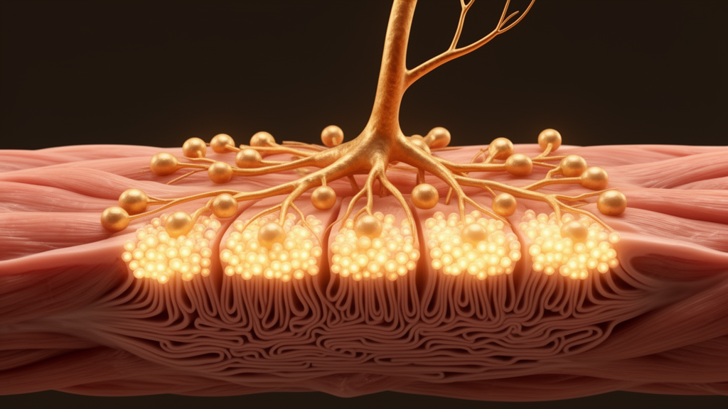
Researchers at the University of Nebraska Medical Center, writing in Frontiers in Bioengineering and Biotechnology, focused on the neuromuscular junction, the specialized synapse where motor neurons command skeletal muscle fibers to contract. Every voluntary movement depends on this microscopic relay. The motor neuron releases the neurotransmitter acetylcholine, which binds to densely packed nicotinic acetylcholine receptors clustered on the muscle membrane at a region called the motor end plate. This binding generates a local depolarization known as the endplate potential, and in healthy tissue a single endplate potential is sufficient to trigger an action potential and a full muscle contraction. The system is built with a large safety margin: the excess of receptors packed into each cluster ensures that every nerve impulse reliably produces movement.
That receptor packing is not accidental. It depends on a cytoplasmic scaffolding protein called rapsyn, which binds to nicotinic acetylcholine receptors and aggregates them at high density into the clusters essential for efficient transmission. Rapsyn does not control how many receptors the muscle produces, but it stabilizes them on the postsynaptic membrane and extends their half-life, slowing their metabolic degradation. In mice genetically engineered to lack rapsyn, motor axons can still form presynaptic nerve terminals and receptors still concentrate at synaptic sites, yet the postsynaptic specialization fails to develop and synaptic activity collapses. Rapsyn, in other words, is the molecular glue that holds the receiving end of the synapse together.
The Nebraska team, led by Yu-Long Li with Huiyin Tu as first author, asked whether tourniquet-induced ischemia-reperfusion injury depletes rapsyn and thereby dismantles the receptor clusters. They used male C57BL/6 mice and placed an orthodontic rubber band at the hip joint of one hindlimb under anesthesia, blocking blood flow for three hours before releasing it to allow six weeks of reperfusion. Blood flow was verified with laser Doppler speckle imaging. The choice of a rubber band rather than a pneumatic cuff was deliberate: the model’s stability depends on consistent limb dimensions, which is also why the researchers used only male mice, since matching males and females for both age and body weight proved impossible with this technique. All animals and tissue samples were blinded during experimentation and data acquisition until statistical analysis.
The results were striking. After six weeks, the neuromuscular junctions in the tourniquet-injured gastrocnemius muscle were severely fragmented, with rapsyn expression markedly reduced and rapsyn itself displaying the same broken pattern as the receptor clusters it normally organizes. In sham-operated mice, only 3.8 percent of junctions were fragmented; in injured mice, the figure soared to 74.1 percent. The total area of each receptor cluster was unchanged, but the area of each individual fragment shrank from roughly 313 square micrometers to about 89, meaning the once-continuous receptor field had shattered into small disconnected pieces. Function followed structure. Endplate potential amplitudes fell from 27.6 millivolts in sham animals to 18.2 millivolts after injury, and electrically stimulated muscle contraction was reduced at every stimulation frequency tested. On a rotarod test, in which mice must stay on an accelerating rotating rod, injured animals fell sooner and at lower speeds than their sham counterparts.
The pivotal experiment came next. Two weeks into reperfusion, once the acutely damaged muscle fibers had been cleared and new myotubes formed, the researchers injected an adenovirus carrying the mouse rapsyn gene directly into the injured gastrocnemius muscle. The transfection was efficient, reaching about 71 percent of muscle cross-section, and raised rapsyn protein levels nearly threefold. The effect on the synapse was dramatic: fragmented junctions dropped from 74.7 percent to 38.2 percent, the area of each receptor fragment recovered substantially, and endplate potential amplitudes rose significantly. Muscle contraction improved at low-frequency stimulation, and the rotarod trajectory trended upward, although the latter improvement did not reach statistical significance. Notably, motor nerve innervation and nerve terminal occupancy at the junction were unaffected by the treatment, indicating the virus was acting on the muscle side of the synapse rather than the nerve.
To confirm that rapsyn depletion is genuinely causal rather than merely correlated, the team ran the experiment in reverse. They injected a lentivirus delivering rapsyn shRNA into the gastrocnemius muscles of normal mice, knocking down the protein without any injury at all. The result mimicked the tourniquet phenotype: receptor clusters fragmented, junctions broke apart, endplate potentials shrank, and muscle contractile function declined, with the strongest impairment appearing during high-frequency nerve stimulation. A scrambled shRNA control had no effect. Together, the overexpression and knockdown data establish rapsyn as a necessary and sufficient regulator of receptor cluster integrity in this injury context.
An intriguing frequency-dependent pattern emerged from the electrophysiology. Rapsyn overexpression rescued contraction evoked by 50-hertz stimulation but not by 100 or 150 hertz, while rapsyn knockdown in healthy muscle spared low-frequency contraction but significantly impaired high-frequency responses. The researchers interpret this to mean that an intact junctional architecture becomes especially critical when the nervous system demands rapid, sustained firing. During emergencies, muscles are called upon to generate force at high frequencies, which may explain why junctional fragmentation after tourniquet injury is so disabling even when some low-frequency capacity survives.
Why does rapsyn disappear after ischemia-reperfusion? The mechanisms remain unresolved, but the authors outline two plausible routes. Prior work has shown that Wnt3a-activated beta-catenin signaling suppresses rapsyn expression and inhibits receptor clustering, and the team’s own pilot data indicate that tourniquet injury raises beta-catenin protein in skeletal muscle, suggesting the injury may be switching on a pathway that actively silences the rapsyn gene. Alternatively, rapsyn protein might be degraded through ubiquitination, the cellular tagging system that marks proteins for destruction. Distinguishing between transcriptional suppression and protein degradation will be essential for designing drugs that act upstream of rapsyn itself.
The study has honest limitations. Rapsyn overexpression was confined to one muscle rather than the whole injured limb, which may explain why the rotarod gains, which depend on coordinated activity of many muscle groups, fell short of significance. The adenoviral vector was not exclusively restricted to muscle fibers, and although rapsyn expression appeared in motor axons, it altered neither innervation nor terminal occupancy. Full recovery of the junction, the authors emphasize, likely requires coordination among the tripartite synapse’s three cellular partners, presynaptic neurons, postsynaptic muscle fibers, and Schwann cells, along with other organizing proteins such as Lrp4, Dok7, MuSK, and dystrophin, which their earlier work showed also shifts away from the muscle membrane after tourniquet application. Even so, the finding that boosting a single scaffolding protein can partially rebuild a shattered synapse and restore measurable muscle function represents a genuine step toward a targeted therapy. Unlike dexamethasone, which the same group previously showed helps junctional recovery but cannot be used long-term because of severe side effects, rapsyn acts locally and mechanistically at the site of injury. For the soldiers, accident victims, and surgical patients whose limbs bear the hidden cost of tourniquets, that specificity could eventually make the difference between a limb that survives and one that thrives.
Subject of Research: Rapsyn overexpression improves neuromuscular junction and skeletal muscle function after tourniquet-induced mouse hindlimb ischemia-reperfusion injuries
Article Title: Rapsyn overexpression improves neuromuscular junction and skeletal muscle function after tourniquet-induced mouse hindlimb ischemia-reperfusion injuries
Article References: Tu, H., Adu-Agyekum, E., Barksdale, A. N., Wadman, M. C., & Li, Y.-L. (2026). Rapsyn overexpression improves neuromuscular junction and skeletal muscle function after tourniquet-induced mouse hindlimb ischemia-reperfusion injuries. Frontiers in Bioengineering and Biotechnology, 14, Article 1906715. https://doi.org/10.3389/fbioe.2026.1906715
Image Credits: AI Generated
DOI: 10.3389/fbioe.2026.1906715
Keywords: neuromuscular junction, rapsyn, tourniquet, ischemia-reperfusion, skeletal muscle, gene therapy, acetylcholine receptors, regenerative medicine, overexpression, improves, neuromuscular, junction
News Source: Gregory Coleman. (October 15, 2026). Rapsyn Overexpression Restores Neuromuscular Junctions After Tourniquet Injuries. Scienmag.