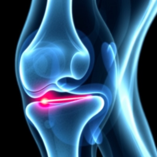
In recent advancements in pediatric radiology, a groundbreaking study has emerged, shedding light on the critical role that history plays in diagnosing foreign body-induced knee synovitis. This condition, while often overlooked, can have significant implications for children’s health if not accurately identified and addressed promptly. For radiologists, the importance of a thorough patient history is underscored, as it can drastically alter diagnostic pathways and therapeutic approaches.
The study conducted by Vassalou et al. marks a pivotal contribution to our understanding of how foreign bodies can influence joint health, particularly in the knee. In children, such occurrences may stem from various activities engaging with small objects, making it imperative for medical professionals to be alert to these cases. The research reinforces the idea that historical context provided by parents or guardians can reveal nuances in a child’s behavior and history that crucially inform diagnosis and treatment.
.adsslot_tWy8jpXA20{ width:728px !important; height:90px !important; }
@media (max-width:1199px) { .adsslot_tWy8jpXA20{ width:468px !important; height:60px !important; } }
@media (max-width:767px) { .adsslot_tWy8jpXA20{ width:320px !important; height:50px !important; } }
ADVERTISEMENT
The radiographic assessment of knee joint conditions requires advanced skills and experience. Radiologists are often the unsung heroes in this diagnosis process. They must be well-versed in potential signs of foreign body presence within the joint space, which may include specific types of effusion or unusual foreign body artifacts on imaging studies. In this regard, the study highlights the significance of imaging modalities such as MRI and ultrasound when it comes to diagnosing foreign body-induced conditions in pediatric patients.
Moreover, the findings advocate for radiologists to engage more actively with the clinical team, facilitating a comprehensive history-taking process. When a parent reports a child’s recent injury or the possibility of foreign body ingestion, these details should prompt an immediate review of the patient’s imaging studies. The studies can be greatly influenced by what a child may or may not have encountered in their environment, and the careful analysis of this information could lead to early intervention and potentially avoid severe complications down the line.
Further, the implications of neglecting patient history can have dire consequences. Delayed diagnosis time can lead to chronic synovitis or even permanent joint damage in young, developing knees. Radiologists, therefore, must prioritize detailed communication with not only referring physicians but also with patients’ families to ascertain crucial details that might seem minor but hold substantial weight in diagnostics.
This research also opens the door for raising public awareness regarding pediatric knee injuries associated with foreign bodies. It stresses the need for guardians to be vigilant about their children’s activities and the potential harmful exposure to small, dangerous materials. Educational initiatives could empower caregivers to recognize symptoms associated with knee synovitis, enabling them to seek prompt medical care when necessary.
In the pursuit of improving diagnostic accuracy, the question arises: how can healthcare systems incorporate structured history-taking into pediatric assessments more effectively? One avenue may involve the development of standardized questionnaires that guide families through pertinent historical details relevant to the child’s condition. This could streamline the communication between families and healthcare providers, ensuring no critical detail is overlooked.
Furthermore, embracing technological advancements in imaging and data collection could also elevate the standard of care. Imagine a scenario in which, during the initial visit to a clinician, a digital tool helps in gathering and synthesizing a patient’s medical history alongside their imaging studies in real time. Such innovations could proactively enhance the diagnostic process in radiology.
As the study by Vassalou and colleagues highlights the critical relationship between history-taking and imaging, it also offers insights into future research directions. More extensive studies could explore the prevalence of foreign body-induced synovitis in various demographics, further helping to inform best practices in pediatric patient management. Understanding how these injuries present across different populations could yield valuable data, leading to refined diagnostic protocols.
In conclusion, the emerging understanding of foreign body-induced knee synovitis emphasizes the multi-faceted role of the radiologist, highlighting how essential a comprehensive patient history is to accurate diagnosis. Engaging with families, utilizing advanced imaging techniques, and advocating for public awareness are all critical steps that can ensure children receive the best care possible. The study serves as a call to action for the medical community to refine diagnostic strategies and ultimately improve patient outcomes in pediatric care.
By focusing on history as a vital component of effective diagnosis in knee synovitis, there lies a promise of better health trajectories for children faced with this often-misunderstood condition. As more research emerges, we can expect to see shifts in diagnostic practices that prioritize history and creativity in imagery interpretation, forever altering the landscape of pediatric radiology.
Subject of Research: Foreign body-induced knee synovitis and the importance of patient history in diagnosis.
Article Title: Foreign body-induced knee synovitis: importance of history by the radiologist.
Article References:
Vassalou, E., Dimitriou, R., Perdikogianni, C. et al. Foreign body-induced knee synovitis: importance of history by the radiologist.
Pediatr Radiol (2025). https://doi.org/10.1007/s00247-025-06339-7
Image Credits: AI Generated
DOI: https://doi.org/10.1007/s00247-025-06339-7
Keywords: knee synovitis, pediatric radiology, foreign bodies, patient history, diagnostic imaging
Tags: challenges of diagnosing knee synovitisdiagnostic pathways for knee conditionsforeign body-induced knee inflammationimplications of untreated knee synovitisimportance of patient history in diagnosisjoint health in childrenmanaging foreign body cases in pediatricsnuances in children’s behavior and health historypediatric radiology advancementsradiologist role in pediatric knee synovitissignificance of thorough medical history in radiologytherapeutic approaches for synovitis

