Researchers at the prestigious University of Adelaide have achieved a groundbreaking milestone in the field of biological imaging with their innovative approach to visualizing live embryos. By utilizing specialized cameras that are originally designed for quantum measurements, the research team has embarked on a transformative journey into the microscopic world, capturing embryos in ways previously thought unattainable. This quantum-enhanced technology allows scientists to investigate dynamic biological processes with unprecedented clarity while minimizing the potential for damage that traditional methods may cause.
The development of this imaging technique arises from the efforts of the Centre of Light for Life at the University of Adelaide. Under the guidance of Director Professor Kishan Dholakia, a leading expert in the field, the team set out to explore how cutting-edge camera technology can enhance the study of life sciences. These advanced cameras are unparalleled in their ability to count individual photons—the basic units of light—at each pixel, thus allowing researchers to examine biological specimens under conditions that replicate their natural environments.
Damaging living cells during imaging has long been a concern for biologists; however, with the advent of this new technology, the potential for harm is significantly reduced. Professor Dholakia states that gentle illumination with low levels of light is essential for accurately understanding living biological processes. “Damage from illumination is a real concern which can often be overlooked. Using the lowest level of light possible, together with these very sensitive cameras is important for understanding biology in live and developing cells,” he emphasized. This approach not only promotes the integrity of living tissues but also opens new dimensions for researchers studying embryonic development.
The research team, comprised of brilliant minds including Zane Peterkovic, Dr. Avinash Upadhya, Ramses Bautista Gonzalez, Dr. Megan Lim, Dr. Chris Perrella, Admir Bajraktarevic, and Associate Professor Kylie Dunning from the Robinson Research Institute, conducted pre-clinical trials to test the efficacy of this innovative imaging technology. Their findings, which were published in the esteemed journal APL: Photonics, reveal a promising future for this application in clinical settings, particularly in the realms of in vitro fertilization (IVF) and reproductive biology.
One of the remarkable outcomes of this research is the realization that fundamental concepts of physics, such as quantum mechanics, can serve as a powerful ally in enhancing digital imaging technology. Lead author and PhD student Zane Peterkovic notes, “Digital camera technology has advanced to the point where fundamental physics concepts like quantum mechanics become important and relevant,” emphasizing the unique intersection of physics and biological sciences. This blend of disciplines fosters deeper insights, ultimately contributing to broader implications for clinical practices and advancements in reproductive health.
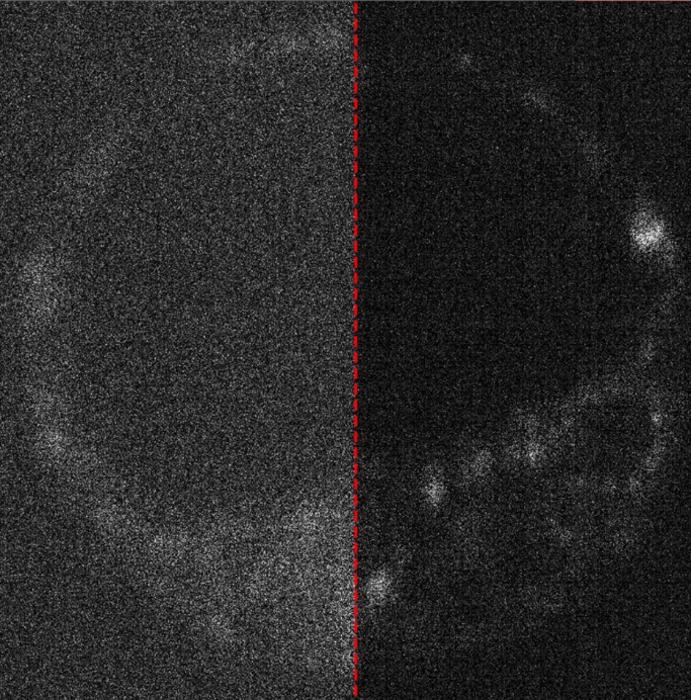
In illuminating biological specimens, natural compounds within cells exhibit fluorescence, revealing substantial insights into their physiological properties. However, capturing these faint signals presents an ongoing challenge. Mr. Peterkovic explained that while the potential for bioluminescence is significant, the emitted signals are often weak. He expressed enthusiasm about applying quantum cameras to harness this delicate fluorescence: “It’s exciting to apply these quantum cameras and use it to get the most out of our microscopes.”
The endeavor involved developing a comprehensive methodology to compare image quality across varying camera technologies effectively. The intricate analysis required collaboration across a wide spectrum of expertise, ranging from optics and biology to laser physics and microscopy techniques. This multidisciplinary approach not only enhances the quality of imaging but also encourages the integration of advanced analytical methods.
The innovation doesn’t stop there; the team investigated how artificial intelligence could be employed to reduce noise from captured images. This static interference is a common challenge when cameras struggle to collect sufficient light. Mr. Peterkovic remarked, “These steps go beyond just putting the camera in the microscope to take pictures,” underscoring the depth of complexity involved in optimizing imaging techniques. Their work seeks not only to advance imaging technology but also to refine the interpretation of the biological data obtained.
Moving forward, the research team sees promising opportunities in the realm of quantum imaging, where manipulating quantum states of light could provide even deeper insights into biological samples. The potential for such advancements could revolutionize the way researchers perceive and understand living systems, pushing the boundaries of current scientific knowledge.
The impact of this research is not limited to academia; industry implications are also significant. By improving the imaging of live embryos, the findings may foster new pathways for clinical applications, especially in reproductive medicine. This could mean better outcomes for patients undergoing IVF treatments, as well as a more profound understanding of developmental biology as a whole.
Acknowledging the support received for this groundbreaking work, Professor Dholakia noted that funding from the Australian Research Council played a crucial role in facilitating the research project. Such collaborations highlight the importance of harnessing resources for scientific endeavors that promise to change the landscape of healthcare and biological research.
In summary, the University of Adelaide’s innovative approach to imaging live embryos using quantum measurement cameras marks a significant advancement in life sciences. By effectively combining the principles of quantum mechanics with cutting-edge imaging technology, the research team has opened new avenues for understanding the complexities of biological processes. As they continue to explore the true potential of these findings, the possibility of transformative breakthroughs in clinical practice and reproductive success becomes increasingly tangible.
Subject of Research: Cells
Article Title: Optimizing image capture for low-light widefield quantitative fluorescence microscopy
News Publication Date: 12-Mar-2025
Web References: DOI Link
References: APL: Photonics Journal
Image Credits: University of Adelaide
Keywords: Quantum imaging, low-light microscopy, biological imaging, fluorescence, IVF, embryology, Australian Research Council, optical technology, laser physics, artificial intelligence, reproductive biology.
Tags: biological imaging advancementsCentre of Light for Lifedynamic biological processesenhancing life sciences research.innovative microscopy techniqueslive embryo visualizationminimizing cell damage in imagingphoton-counting camerasProfessor Kishan DholakiaQuantum imaging technologytransformative imaging methodsUniversity of Adelaide research


