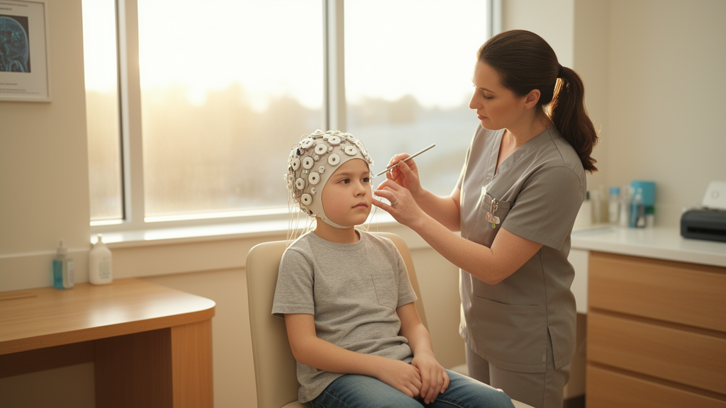
In awake children without neurological disorders or neuroactive medications, quantitative electroencephalography reveals pronounced age-dependent distributions across total power, individual frequency spectra, and the alpha/delta ratio. Continuous full-channel electroencephalography remains the established gold standard for evaluating electrocortical function in pediatric patients facing high risks of acute cerebral injury during critical illness. However, full-channel monitoring and timely real-time review by clinical neurophysiology experts are frequently unavailable across hospital units. To overcome these logistical barriers and prompt timely targeted examinations of raw electroencephalograms, quantitative electroencephalography transforms complex electrical signals into numerical and visual trends that summarize cerebral rhythm, amplitude, frequency, and symmetry at the bedside.
To establish baseline physiological distributions for electrode configurations routinely utilized in pediatric intensive care units, researchers conducted a post-hoc analysis evaluating 450 unremarkable electroencephalograms recorded from individual awake pediatric patients aged between 1 month and 17 years. The underlying cohort distribution was skewed toward older children, with males accounting for 52.7 percent of the participants. The median electroencephalogram recording time was 17 minutes, with an interquartile range spanning 15 to 18 minutes. All included tracings were derived from children without known neurological pathologies, and recordings were performed prior to therapy initiation rather than during episodes of acute critical illness.
The study cohort drew from diverse clinical indications where baseline neurophysiological assessments were standard protocol. At the study center, which houses a comprehensive pediatric oncology department, all patients scheduled for chemotherapy underwent pre-treatment electroencephalography to document baseline neurological function. This identical surveillance protocol applied to pediatric candidates undergoing solid organ or bone marrow transplantation, irrespective of their primary diagnosis. Other evaluations involved diagnostic workups for suspected neurological conditions where subsequent testing definitively excluded any neurological diagnosis both before and after recording. In cases involving acquired brain injuries, only pre-insult tracings met eligibility criteria, while central nervous system involvement in oncology patients was systematically ruled out using neuroimaging or lumbar punctures. Furthermore, patient histories were screened to ensure the complete absence of neuroactive substances at the time of monitoring.
Full-channel recordings adhered to the standard international 10–20 system and were reviewed and verified as unremarkable by a board-certified pediatric neurologist certified in electroencephalography and epileptology by the German Society for Epileptology. Signal processing was performed using Persyst 14 software, focusing on derivations commonly applied in continuous reduced-channel intensive care monitoring: central-parietal channels C3-P3 and C4-P4, as well as the frontal lead Fp1-Fp2. While central-parietal pairs represent standard configurations for amplitude-integrated electroencephalography, the frontal derivation was included because frontal electrode placements may serve as the sole accessible recording sites in patients presenting with traumatic head injuries or following neurosurgical interventions.
Spectral power in microvolts squared was exported in 0.5-hertz bins spanning frequencies from 0 to 20 hertz alongside continuous metadata evaluating signal quality and artifact intensity. Epochs exhibiting artifact intensity exceeding 20 were excluded from analytical pipelines. Band boundaries followed standardized limits, defining delta activity as 0.5 to 3.5 hertz, theta as 3.5 to 7.5 hertz, alpha as 7.5 to 12.5 hertz, and beta as 12.5 to 30 hertz. Total power was computed by summing the power across all frequency bands, and median values were derived for each recording tracing. Relative power was quantified as a percentage of total power, while the alpha/delta ratio was calculated directly by dividing relative alpha power by relative delta power. For percentile calculations and visualization across established developmental age groups, C3-P3 and C4-P4 derivations were consolidated into a unified central-parietal channel.
The quantitative results demonstrated that total electrical power decreased progressively as child age increased, a pattern evident across both central-parietal channels and frontal leads. Overall total power measurements in central-parietal leads remained consistently lower than the amplitudes recorded from the frontal derivation across developmental groups. Spectral analysis revealed that relative power distributions varied notably between specific age brackets and recording channels. In central-parietal derivations, delta power dominated electrocortical activity in children younger than 6 years of age, representing 49.1 to 39.3 percent of relative power at the 50th percentile, before declining to 32.7 to 27.6 percent among older subjects aged 6 to 17 years.
Theta band activity in central-parietal channels was most prominent among children younger than 1 year, where it accounted for a median relative power of 23.1 percent, subsequently increasing to 25.7 percent in the 2- to 5-year-old cohort. Conversely, the frontal Fp1-Fp2 channel exhibited a markedly different profile, with delta activity remaining the dominant frequency band across every developmental stage, registering at 50.4 percent in infants under 1 year and 43.5 percent in adolescents aged 14 to 17 years. In contrast to the shifting central-parietal dynamics, relative alpha, beta, and theta power remained largely stable across all evaluated age groups within the frontal derivation.
The alpha/delta ratio within central-parietal channels showed extensive developmental variation across childhood. In children younger than 1 year, the central-parietal alpha/delta ratio ranged between 0.1 and 0.4 from the 5th to the 95th percentiles. This ratio rose continuously with advancing maturity, ultimately reaching a distribution of 0.5 to 2.3 among adolescents aged 14 to 17 years. By comparison, frontal derivations demonstrated far less developmental variation, with the alpha/delta ratio remaining tightly constrained between 0.1 and 0.6 across all pediatric age cohorts.
These electrophysiological findings emphasize the physiological principles underlying cerebral maturation. Lower frequencies like delta and theta rhythms originate within thalamic structures and deep cortical layers II through VI, whereas faster alpha frequencies emerge predominantly from cortical layers IV and V. In clinical intensive care environments, alterations in spectral power and alpha/delta ratios have shown utility in identifying secondary complications, including delayed cerebral ischemia, stroke, traumatic brain injuries, and emergence delirium. Because spectral shifts can precede overt clinical deterioration by several hours, establishing precise developmental reference ranges for practical reduced-electrode arrays is essential to prevent normal developmental patterns from being misinterpreted as neurological decline.
The authors highlighted several critical boundaries regarding the scope and generalizability of the findings. All data were acquired exclusively from awake, neurologically unremarkable pediatric patients who were free of centrally active medications, representing physiological conditions that rarely match the sedated, critically ill status of pediatric intensive care patients. Consequently, the observed reference values do not reflect electrophysiological modifications induced by natural sleep stages or sedative infusions. Additionally, because the analysis was restricted to specific central-parietal and frontal derivations, the findings cannot be directly extrapolated to other montage configurations. Future studies will be required to define quantitative electroencephalography distributions under sleep and sedative conditions in critically ill children.
The establishment of normative baseline ranges addresses an important void in neurocritical literature, where detailed analyses spanning individual alpha, beta, theta, and delta frequencies have been remarkably scarce compared to isolated assessments of the alpha/delta ratio. Prior investigations demonstrate that distinct spectral band behaviors carry meaningful prognostic utility across diverse acute conditions. For instance, researchers identified that a power reduction exceeding 40 percent persisting for more than 5 hours within the alpha band and over 6 hours within the theta band established an optimal diagnostic threshold for identifying delayed cerebral ischemia, defined by clinical symptoms or neuroimaging verification on computed tomography or magnetic resonance imaging. Furthermore, electrophysiological shifts across theta and delta bands have been documented to precede overt clinical deterioration in pediatric patients by several hours. In mechanical ventilation contexts, relative power stability across all four frequency bands has been linked to successful patient extubation, whereas elevated volatility in frontal beta activity predicted extubation failure, demonstrating the clinical necessity of tracking each individual frequency band.
The developmental trajectories observed in the current pediatric cohort also provide essential nuance for interpreting pathological declines in the alpha/delta ratio. Reductions in this metric, paired with increases in relative delta power, are well-documented hallmarks of compromised cerebral perfusion following subarachnoid hemorrhage and ischemic stroke in adult populations. Previous pediatric research investigating 49 patients undergoing extracorporeal membrane oxygenation similarly demonstrated substantial alterations in the alpha/delta ratio as an indicator of cerebral perfusion changes; however, that comparison cohort remained young and lacked developmental stratification. By demonstrating that the ratio naturally increases with age in healthy children, the present data indicate that pathological thresholds must be evaluated against age-adjusted normative baselines. Additionally, the observed central-parietal patterns contrast with standard full-channel recordings, where the dominant background rhythm in awake children is typically an occipital alpha rhythm that increases with age while theta power remains largely age-independent.
The pronounced electrophysiological differences documented between frontal and central-parietal derivations also illuminate technical considerations inherent to bedside monitoring. Frontal channels exhibited notably higher total power alongside constrained ratio variation across age groups, a divergence the investigators attributed partly to physiological artifacts generated by cranial muscle activity. Such myogenic interference is known to be particularly prominent in frontal electrode placements, presenting a significant factor in cohorts comprising fully awake subjects. All raw data transformations in the investigation relied on Fast Fourier Transformation to extract frequency bins, with subsequent data preparation and statistical computations executed within Jupyter Lab using Python 3.13. By defining reference distributions across these specialized configurations, the dataset establishes an empirical foundation for recognizing acute neurophysiological deviations in intensive care environments, with underlying research data made available by the study authors upon reasonable request.
Subject of Research: Age-specific quantitative electroencephalography reference values in pediatric patients
Article Title: Age-specific distributions of total power, frequency bands, and alpha/delta ratio in quantitative EEG
Article References: Greve, S., Brensing, P., Paul, L., Dohna-Schwake, C., Rummeni, F., Felderhoff-Mueser, U., & Bruns, N. (2025). Age-specific distributions of total power, frequency bands, and alpha/delta ratio in quantitative EEG. Intensive Care Medicine – Paediatric and Neonatal, 3(1), Article 39. https://doi.org/10.1007/s44253-025-00092-3
Image Credits: AI Generated
DOI: 10.1007/s44253-025-00092-3
Keywords: quantitative electroencephalography, pediatric intensive care, alpha/delta ratio, total power, delta frequency, theta frequency, alpha frequency, beta frequency, reduced electrode sets, electrocortical function, central-parietal channels, frontal leads, neuromonitoring, brain development, spectral power, fast fourier transform
News Source: Ophelia Keating. (November 20, 2026). Quantitative EEG Power and Alpha-Delta Ratios Shift With Age in Children. Scienmag.