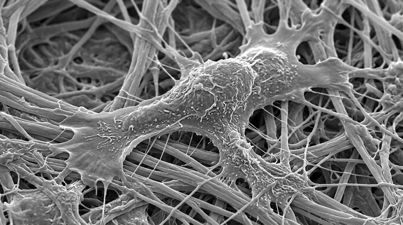
Researchers have identified a specific molecular mechanism by which the enzyme PTGS1 regulates macrophage polarization, thereby influencing inflammation and oxidative stress in chronic prostatitis. Published in the Journal of Cellular and Molecular Medicine, this study demonstrates that silencing PTGS1 promotes the shift of macrophages toward an anti-inflammatory M2 phenotype. This shift is mediated through the nuclear translocation of the protein PPARγ, which subsequently reduces tissue injury in a mouse model of experimental autoimmune prostatitis (EAP). The findings offer a new perspective on the role of prostaglandin-endoperoxide synthase 1 (PTGS1) in immune regulation, moving beyond its traditional characterization as a constitutive enzyme involved in homeostatic prostaglandin production.
Chronic prostatitis/chronic pelvic pain syndrome (CP/CPPS) is a common urological disorder affecting men across various age groups, with an estimated lifetime prevalence of 9.3%. The condition is characterized by persistent pelvic pain and lower urinary tract symptoms, including urinary difficulty, urgency, and frequency. Because CP/CPPS is multifactorial and often resistant to current treatments, understanding its underlying inflammatory and immune mechanisms is critical for developing more effective therapeutic strategies. Immune dysfunction is a key pathological basis for the onset and progression of the disease, with macrophages playing a central role due to their ability to polarize into pro-inflammatory M1-like or anti-inflammatory M2-like phenotypes.
The study began by analyzing three Gene Expression Omnibus (GEO) datasets related to IL-4-induced M2 macrophage polarization: GSE151015, GSE183074, and GSE208394. A Venn diagram analysis of these datasets identified 123 overlapping differentially expressed genes, among which PTGS1 was selected for further validation due to its known involvement in prostaglandin synthesis and inflammatory regulation. When researchers treated immortalized bone marrow-derived macrophage (iBMDM) cells with IL-4 to induce M2 polarization, quantitative PCR confirmed the upregulation of PTGS1 transcripts. This increase in PTGS1 protein expression was further corroborated by immunofluorescence and western blotting, indicating that PTGS1 is dynamically regulated during the M2 polarization process.
To investigate the functional consequences of PTGS1 expression, the researchers used lentivirus-mediated shRNA to achieve efficient PTGS1 knockdown in macrophages. Loss of PTGS1 led to a marked induction of M2-specific markers, including Arg1, YM1, Fizz, CD206, and IL-10, as evidenced by western blotting and RT-qPCR. Flow cytometry showed that PTGS1 depletion increased the proportion of CD206+ M2 macrophages under IL-4 stimulation. Functionally, PTGS1 knockdown augmented arginase activity while diminishing the secretion of nitric oxide (NO) and reactive oxygen species (ROS). These results suggest that PTGS1 acts as a feedback regulatory checkpoint that restrains the full activation of the M2 polarization program.
RNA sequencing of control and PTGS1-silenced macrophages after IL-4 stimulation revealed widespread transcriptomic alterations, with gene set enrichment analysis (GSEA) showing significant enrichment of gene sets associated with nuclear receptor signalling. Co-immunoprecipitation experiments demonstrated that PTGS1 specifically interacts with PPARγ, a key regulator of macrophage polarization and inflammatory responses. This interaction was more pronounced in M2 macrophages. Crucially, PTGS1 knockdown reduced cytoplasmic PPARγ levels and promoted its accumulation in the nucleus. This indicates that the PTGS1-PPARγ interaction inhibits PPARγ nuclear translocation, thereby limiting PPARγ-dependent M2-promoting transcriptional activity.
To confirm the dependency of PTGS1-driven polarization on PPARγ, the researchers administered the PPARγ inhibitor GW9662 to PTGS1-knockdown macrophages. This treatment reversed the M2 phenotype, as evidenced by the downregulation of characteristic M2 markers and factors. Western blot analysis showed that the induction of Arg1 and YM1 following PTGS1 silencing was abolished by GW9662, confirming that their expression is PPARγ-dependent. Flow cytometry confirmed a significant decrease in the M2 macrophage proportion upon PPARγ inhibition, while pro-inflammatory mediators NO and ROS were elevated. These findings identify PPARγ as the critical mediator through which PTGS1 regulates M2 macrophage polarization.
In vivo experiments were conducted using a myeloid-specific PTGS1 knockdown EAP mouse model. The EAP model was established through prostate antigen and complete Freund’s adjuvant immunization, confirmed by increased pelvic pain hypersensitivity and histopathological inflammatory infiltration in prostate tissues. PTGS1 silencing led to a significant improvement in disease phenotypes, manifested as a lower frequency of pain responses and diminished inflammatory damage in prostate tissue. At the molecular level, PTGS1 silencing counteracted the EAP-induced cytokine imbalance by lowering IL-1β, IL-6, and TNF-α and elevating IL-10. Flow cytometry revealed that PTGS1 knockdown increased the proportion of CD206+ M2 macrophages and decreased the proportion of M1 macrophages in the spleen.
Immunofluorescence analysis of prostate tissue showed that PTGS1 knockdown reduced the accumulation of F4/80+/iNOS+ pro-inflammatory macrophages. A comprehensive assessment of oxidative stress in these mice revealed that EAP mice exhibited a significant increase in ROS, which was effectively mitigated by PTGS1 silencing. PTGS1 knockdown also reduced EAP-induced cellular apoptosis, as indicated by TUNEL staining, and decreased lipid peroxidation, as shown by 4-HNE immunohistochemistry. Biochemical assays confirmed that PTGS1 silencing normalized elevated serum malondialdehyde (MDA) levels and restored serum superoxide dismutase (SOD) activity. Western blot analysis showed upregulation of the NRF2/HO-1 antioxidant pathway in EAP mice, which was suppressed upon PTGS1 knockdown.
To definitively establish whether PTGS1 functions through PPARγ in vivo, the PPARγ inhibitor GW9662 was administered to myeloid-specific PTGS1 knockdown EAP mice. This treatment abolished the protective effects of PTGS1 silencing, exacerbating prostate tissue inflammation and increasing the frequency of pain responses. The cytokine profile shifted toward a pro-inflammatory state, with elevated levels of IL-1β, IL-6, and TNF-α and a reduction in IL-10. The increase in CD206+ M2 macrophages conferred by PTGS1 knockdown was reversed upon PPARγ inhibition. Furthermore, GW9662 treatment intensified pro-inflammatory macrophage accumulation and oxidative stress, including increased ROS, TUNEL staining, and 4-HNE immunoreactivity, along with elevated serum MDA and decreased SOD activity.
These results confirm that the beneficial effects of PTGS1 silencing on oxidative stress, inflammation, and pain in EAP mice are mediated primarily through the PPARγ pathway. The study clarifies that IL-4-induced PTGS1 upregulation does not indicate that PTGS1 acts as a positive driver of M2 polarization. Instead, the data support a feedback regulatory model in which PTGS1 is induced during IL-4-mediated macrophage polarization and subsequently restrains excessive PPARγ nuclear translocation by interacting with PPARγ in the cytoplasm. By removing this regulatory checkpoint, PTGS1 knockdown facilitates PPARγ nuclear accumulation and strengthens the M2 polarization program, thereby attenuating inflammatory progression and oxidative stress damage.
From a translational perspective, this study provides a mechanistic rationale for targeting macrophage polarization in CP/CPPS. Current management of CP/CPPS remains largely symptom-oriented, including alpha-blockers, anti-inflammatory agents, and antibiotics in selected patients. However, mechanism-directed immunomodulatory strategies remain limited. By identifying PTGS1 as a feedback regulatory checkpoint of PPARγ-dependent macrophage polarization, the study highlights a potential therapeutic entry point for developing macrophage-targeted interventions. The precise molecular details by which PTGS1 regulates PPARγ trafficking and activity remain incompletely elucidated and require further mechanistic exploration. Additionally, PTGS1 expression was not resolved at single-cell resolution within the prostatic immune microenvironment, suggesting that future studies using single-cell RNA sequencing and spatial validation are needed to characterize PTGS1 expression across distinct macrophage subsets.
Despite these limitations, the data clearly underscore the critical role of PTGS1 in the pathogenesis of chronic prostatitis. The identification of the PTGS1-PPARγ axis as a potential immunomodulatory target offers a new direction for research into the treatment of CP/CPPS.
Subject of Research: The role of PTGS1 in regulating macrophage polarization and oxidative stress in chronic prostatitis via the PPARγ pathway.
Article Title: PTGS1 Regulates Macrophage Polarization Through PPARγ Nuclear Translocation to Affect Oxidative Stress Injury in Chronic Prostatitis
Article References: Song, J., Li, W., Han, S., Song, W., Liang, C., Liu, Y., Zhang, K., & Chen, X. (2026). PTGS1 Regulates Macrophage Polarization Through PPARγ Nuclear Translocation to Affect Oxidative Stress Injury in Chronic Prostatitis. Journal of Cellular and Molecular Medicine, 30(21), Article e71397. https://doi.org/10.1111/jcmm.71397
Image Credits: AI Generated
DOI: 10.1111/jcmm.71397
Keywords: PTGS1, Macrophage Polarization, Chronic Prostatitis, PPARγ, Oxidative Stress, M2 Macrophages, Inflammation, Experimental Autoimmune Prostatitis, Immunomodulation, Urology, Regulates, Macrophage
News Source: Drew Townsend. (November 8, 2026). PTGS1 Silencing Boosts M2 Macrophages to Ease Prostatitis. Scienmag.