In the realm of pediatric radiology, the discovery of normal variants in anatomical structures can often lead to confusion for both clinicians and parents alike. In a recent study published in Pediatr Radiol, researchers Zuniga, Lopez, and Simonton have shed light on a previously under-discussed subject: the proximal medial tibial metaphyseal bump. This anatomical feature, often misidentified as a pathological entity, is revealed to be a normal variation in the growing child’s anatomy. This revelation not only enhances our understanding of pediatric bone development but also has profound implications on diagnostic imaging practices.
Understanding the significance of the proximal medial tibial metaphyseal bump requires a basic grasp of tibial anatomy. The tibia, commonly referred to as the shin bone, plays a critical role in weight-bearing and mobility. Its proximal end features an intricate structure designed to provide stability to the knee joint. Among the many nuances of this section of the tibia, the metaphyseal regions are vital. These areas are responsible for the bone’s growth and are characterized by high cellular activity. The bump described in the study is found at the metaphyseal junction where growth plate activity is most pronounced.
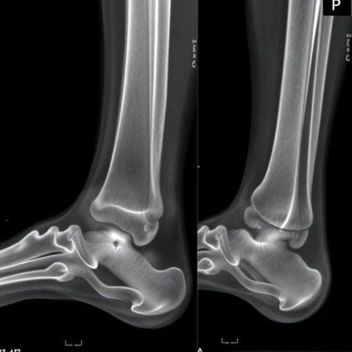
The researchers conducted a thorough analysis on a large sample of pediatric patients, examining both plain radiographs and MRI scans. Any incidental findings of the proximal medial tibial metaphyseal bump were meticulously documented. This approach led to a comprehensive understanding of how prevalent this anatomical variant is in healthy children versus how frequently it is misinterpreted as a sign of pathology, such as osteomyelitis or a fracture. The study found that the bump is not only common but also innocuous, reducing the burden of unnecessary anxiety and further invasive procedures in pediatric patients.
.adsslot_WvYgdKO3Xf{width:728px !important;height:90px !important;}
@media(max-width:1199px){ .adsslot_WvYgdKO3Xf{width:468px !important;height:60px !important;}
}
@media(max-width:767px){ .adsslot_WvYgdKO3Xf{width:320px !important;height:50px !important;}
}
ADVERTISEMENT
In clinical practice, radiologists often face the challenge of differentiating between normal anatomical variants and actual pathologies. The proximal medial tibial metaphyseal bump is an excellent demonstration of this dilemma. The study encourages practitioners to recalibrate their readings of tibial radiographs. By acknowledging this feature as a normal variant, clinicians can avoid unnecessary follow-up imaging or consultations, ultimately leading to more streamlined and patient-centered care. This shift in understanding is pivotal for fostering better diagnostic accuracy.
Moreover, the researchers posed an interesting question: what role does age play in the prominence of the proximal medial tibial metaphyseal bump? As children grow, their bones undergo significant remodeling. The study showed that while the bump is consistently observable in various age groups, its size and shape may change, reflecting the individual growth patterns of each child. This finding underscores the dynamic nature of pediatric bone development and reinforces the idea that what is perceived as a normal variant must be understood in the context of developmental stages.
In the landscape of pediatric medicine, early and accurate diagnosis can significantly influence treatment outcomes. With the identification of the proximal medial tibial metaphyseal bump as a benign anatomical variant, we can reduce the risk of misdiagnosis. This enhancement in diagnostic proficiency ensures that clinicians remain focused on genuine pathological conditions that may require intervention, rather than being sidetracked by features that are simply a part of normal development.
As this research continues to circulate among pediatricians and radiologists, it is likely to influence the training of incoming professionals. Incorporating this knowledge into medical education can help mold a new generation of clinicians who approach pediatric imaging with a critical and informed perspective. This will ensure that they are better equipped to navigate the complexities of childhood anatomy and physiology.
In summary, the emergence of the proximal medial tibial metaphyseal bump as a recognized normal variant is a game-changer in the field of pediatric imaging. This study not only highlights the necessity of differentiating between normal anatomy and disease but also exemplifies the ongoing need for research that explores the complexities of pediatric development. As we continue to learn more about the human body’s variances, it paves the way for more tailored, precise, and compassionate care.
In the end, this new insight also has implications for future studies. With the identification of the proximal medial tibial metaphyseal bump as a benign feature, researchers are encouraged to continue exploring other potential variants in the pediatric population. Such knowledge could pave the way for identifying and understanding other anatomical variations across different ethnicities and populations, thereby enriching the field of pediatric medicine even further.
The collective findings from Zuniga et al. serve to remind medical professionals that the human body, particularly in its developmental phases, is a complex and varied masterpiece. Recognizing the nuances of normal growth is essential in fostering a more significant understanding of pediatric healthcare.
Subject of Research: Proximal medial tibial metaphyseal bump as a normal variant in pediatric patients.
Article Title: Proximal medial tibial metaphyseal bump—a normal variant.
Article References: Zuniga, B., Lopez, N., Simonton, K. et al. Proximal medial tibial metaphyseal bump—a normal variant. Pediatr Radiol (2025). https://doi.org/10.1007/s00247-025-06361-9
Image Credits: AI Generated
DOI: https://doi.org/10.1007/s00247-025-06361-9
Keywords: pediatric radiology, tibial anatomy, normal variants, metaphyseal bump, diagnostic imaging, pediatric medicine, bone development.
Tags: anatomical variants confusion in cliniciansbone development in growing childrendiagnostic challenges in pediatric radiologygrowth plate activity in childrenimplications for diagnostic imagingnormal anatomical variations in pediatricspediatric bone anatomy misconceptionspediatric radiology normal variantsproximal medial tibial metaphyseal bumptibial anatomy in childrentibial metaphysis featuresunderstanding knee joint stability
