A $650 Open-Source Scanner Could Bring Real-Time, Radiation-Free Imaging to Space and Remote Clinics
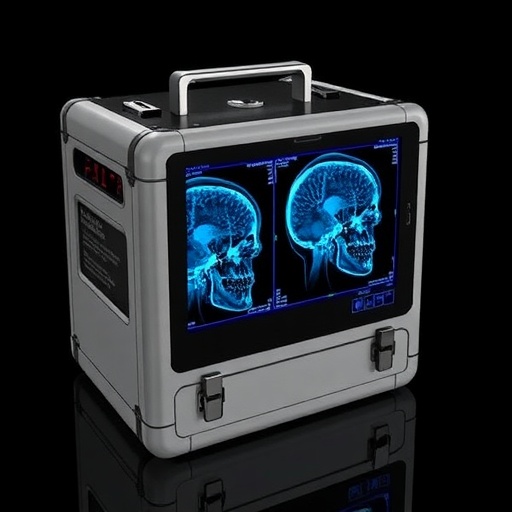
A shoebox-sized medical imaging system built from commercially available electronics could make continuous, radiation-free monitoring practical in places where MRI and computed tomography are impossible—including remote clinics, disaster zones and, eventually, spacecraft. Researchers have developed a 64-channel electrical impedance tomography (EIT) data-acquisition platform that costs about $650 in hardware, fits within a 10 × 8 × 8-centimeter stack and is openly documented so that other laboratories can reproduce or modify it. The device does not produce conventional anatomical images with the sharpness of an X-ray or MRI scan. Instead, it measures how biological tissues resist and store electrical energy, then uses those measurements to reconstruct changing maps of electrical properties inside the body. That lower spatial resolution is balanced by portability, low cost, high temporal resolution and the ability to image continuously without ionizing radiation. The prototype was designed as a ground-based demonstration for future aerospace medical systems, where mass, power, storage and crew time are all severely constrained.
Electrical impedance tomography works by surrounding or covering a region of the body with electrodes. The instrument injects a small alternating current through one pair of electrodes and measures the resulting voltages at other pairs. Because tissue impedance is defined by the relationship between voltage and current, the system calculates complex impedance using Ohm’s law, Z = V/I. “Complex” here means that the measurement includes both resistance and reactance: biological tissues can dissipate electrical energy, store it, and alter the phase of an applied signal. The resulting boundary measurements are fed into an inverse mathematical problem to estimate the electrical properties within the body. Air-filled lungs, blood, muscle, fat and injured or tumorous tissue can produce different impedance signatures, and changes in those signatures can reveal respiration, muscle activity, fluid movement, tissue damage or potentially internal bleeding. EIT is therefore not competing directly with MRI or CT for detailed static anatomy; it is aimed at fast functional monitoring, where a stream of less sharply resolved images may be more valuable than an occasional high-resolution scan.
The new instrument tackles a persistent engineering problem in compact EIT systems: shrinking the electronics without sacrificing frequency flexibility, channel count or measurement speed. Large commercial EIT platforms often use parallel measurement circuits, with dedicated electronics for many electrode channels. That architecture is fast, but it increases board size, power consumption and the number of digital connections. The Dartmouth EIT Edge-64 instead uses a semi-parallel design controlled by an Artix-7 field-programmable gate array, or FPGA. An FPGA is a reconfigurable integrated circuit whose logic can be rewritten after manufacture, unlike an application-specific integrated circuit, which is fixed and expensive to redesign. The controller coordinates a voltage source, programmable amplifiers, analog-to-digital converters and multiplexers that select which electrodes inject current or measure voltage. By sharing measurement resources through rapid switching, the system supports 64 electrodes in a compact package while retaining the ability to adapt its operating modes for different imaging applications.
At the heart of the analog front end is a digitally tunable direct-digital-synthesis voltage source. The system can generate either a single sine wave or two synchronized tones, with selectable frequencies spanning approximately 100 hertz to 3 megahertz. This broad range matters because tissue impedance changes with frequency. At lower frequencies, current tends to be restricted by cell membranes and extracellular pathways; at higher frequencies, capacitive membrane effects become less dominant and current can interact differently with intracellular and tissue-level structures. Measuring at multiple frequencies can therefore add contrast between tissue types and pathological states. Two-frequency operation is especially useful because it avoids switching between every frequency and can support frequency-difference EIT in near real time. The design uses lookup tables to match each generated frequency with the appropriate fast Fourier transform bin used for demodulation. A 24-bit DDS provides frequency resolution of about 5.96 hertz when driven by a 100-megahertz clock. Adding a second tone divides the available signal amplitude between the waves, producing an expected signal-to-noise penalty of roughly 3 decibels, but the simultaneous acquisition can substantially reduce timing overhead.
The measurement circuitry was engineered around the difficult electrical environment presented by the human body and electrode interface. Each source path includes a 50-ohm sense resistor and a 10-microfarad DC-blocking capacitor. The capacitor prevents direct current from flowing through the patient, while the resistor enables the instrument to estimate the injected current. Two voltage-pickup channels use OPA2810 operational amplifiers, selected for their high common-mode rejection, rapid slew rate, very high input impedance and low noise. Programmable-gain amplifiers can select gains from 1 to 32, allowing the circuit to handle signals across a wide impedance range. Three 16-bit, 15-megasample-per-second analog-to-digital converters digitize the current and voltage signals. Custom differential drivers shift and condition the signals for the converters, which accept differential inputs centered around 2.048 volts. High-precision voltage references stabilize those levels, helping the FPGA perform repeatable complex demodulation of the amplitude and phase information carried by the measured waveforms.
Electrode selection is managed by 74HC4067 16-channel multiplexers arranged in cascaded groups. The FPGA can designate any electrode as the positive source, negative source or one of two voltage pickups. Four multiplexers are used for each source output and two for each pickup input, creating the 64-channel interface. Multiplexers introduce resistance and capacitance, which can attenuate signals and shift their phase, particularly at high frequencies or when the load impedance is large. The selected chips have a typical on-resistance of about 90 ohms and source/drain capacitance of 45 picofarads. Those imperfections are partly handled by independently measuring the source current in the tetrapolar configuration, in which impedance is calculated from the measured load voltage divided by the measured current. For time-difference imaging, the same signal paths are used for reference and measurement datasets, reducing the impact of channel-to-channel variations. Absolute imaging, by contrast, may require calibration with a known phantom to correct gain and phase errors. The accompanying MATLAB interface can display individual channel responses, frequency sweeps, complex impedance and time-domain signals, making it possible to identify a poorly connected electrode or malfunctioning channel.
The FPGA’s digital architecture is what allows the small instrument to process signals rapidly while remaining reprogrammable. A 1024-point fast Fourier transform extracts the real and imaginary components of the selected frequency bins from the digitized data. The system uses several clock domains: a 100-megahertz system clock, a 125-megahertz clock for the DAC, a 200-megahertz low-voltage differential-signaling link to the ADCs and a 15-megahertz sampling clock for converted data. Moving information safely between these clock rates is a classic digital-design hazard because an asynchronous transition can leave a logic element in an unstable state, a condition known as metastability. The engineers use flip-flop synchronizers, pulse-lengthening circuits and dual-clock first-in-first-out buffers to stabilize individual signals and larger data transfers. Rather than storing every sample, the FPGA summarizes each acquisition by retaining the relevant signal bins, the direct-current bin, the sum of squared spectral magnitudes for signal-to-noise calculations and a dataset index. This reduces data-transfer demands and leaves memory for up to 2,048 datasets, useful for averaging or sequential measurements.
Testing showed that the prototype met or exceeded many of its design goals, although its performance depends strongly on the chosen imaging protocol. Across resistive loads from 10 ohms to 10 kilohms and frequencies from 100 hertz to 3 megahertz, the system achieved impedance precision better than 1 ohm for loads up to 3 kilohms and better than 1.3 ohms for loads up to 10 kilohms. With four-dataset averaging, its signal-to-noise ratio exceeded 80 decibels for loads above 50 ohms between 1 kilohertz and 1 megahertz. Without averaging, the signal-to-noise ratio was typically 60–70 decibels, still within the range useful for EIT reconstruction. The evaluation used discrete electrical loads rather than human subjects, so these results characterize the electronics and do not establish diagnostic performance. Human measurements would introduce electrode-contact variability, motion, biological noise and safety considerations that cannot be reproduced fully with resistors and capacitors. The hardware also needs to comply with medical electrical-safety and electromagnetic-compatibility requirements before clinical or crewed-space use.
Speed is governed by a fundamental tradeoff between frequency coverage, electrode patterns, averaging and signal quality. A complete sweep of 64 predefined frequencies takes about 299 milliseconds for a standard tetrapolar configuration, but most of that time is spent sampling the lower-frequency signals. The lower 32 frequencies, extending from 100 hertz to 24.3 kilohertz, require slow acquisition and take about 294.3 milliseconds; the upper 32 frequencies, from roughly 14 kilohertz to 3 megahertz, require only about 3 milliseconds. In selected configurations, the system can exceed the target of 30 frames per second. A single frequency or dual-tone measurement above 14 kilohertz can support as many as 62 source-and-pickup combinations at that rate, while a comparable low-frequency measurement supports about three combinations. Fourfold averaging improves noise performance but reduces the number of combinations available at 30 frames per second to about 40 in one tested configuration. Sixty-four unique source-and-pickup patterns at frequencies above 14 kilohertz reached approximately 29.2 frames per second, just below the target. These figures illustrate why a future imaging system would need to tailor its acquisition strategy to the physiological process being monitored.
The platform’s most consequential feature may be that its design files are openly available. Schematics, printed-circuit-board manufacturing files, a bill of materials, FPGA bitstreams, VHDL source code and MATLAB software are released through a Zenodo repository under a combination of CERN open-hardware and MIT licenses, with documentation under a Creative Commons license. Other groups can therefore reproduce the two custom circuit boards, alter the FPGA logic, add frequency modes or adapt the electrode interface for new probes without starting from a proprietary design. The Artix-7 used in the prototype is not radiation-hardened, but its logic architecture is intended to be portable to more expensive space-grade FPGA families. That makes the device a proof of concept rather than a ready-to-fly medical scanner. Still, an open, low-power and reconfigurable EIT platform could give researchers a practical foundation for monitoring lung function, fluid shifts, muscle atrophy or injury during spaceflight, while also lowering the barrier to medical imaging in remote and resource-limited settings. Its promise lies not in replacing every scanner, but in making continuous electrical imaging available where conventional machines cannot go.
Subject of Research: Open-source, FPGA-based 64-channel electrical impedance tomography data-acquisition hardware for portable and aerospace medical imaging.
Article Title: A $650 Open-Source Scanner Could Bring Real-Time, Radiation-Free Imaging to Space and Remote Clinics
Article References: Dartmouth EIT Edge-64 design files and documentation, Zenodo repository: https://doi.org/10.5281/zenodo.21359298
Image Credits: AI Generated
DOI: 10.5281/zenodo.21359298
Keywords: electrical impedance tomography, EIT, medical imaging, FPGA, open-source hardware, aerospace medicine, portable diagnostics, radiation-free imaging, bioimpedance, real-time monitoring
Tags: 64-channel EIT data acquisition systemaerospace medical imaging solutionsaffordable portable medical imaging devicesbiomedical impedance tomography for remote clinicscompact high-speed imaging technologyelectrically resistive tissue mappinghigh temporal resolution bioelectrical measurementslow-cost medical imaging systemopen-source EIT hardware platformportable FPGA-based electrical impedance tomographyradiation-free continuous monitoring devicespace and disaster zone medical imaging