Pretherapeutic quantitative iodine-131 single-photon emission computed tomography combined with computed tomography reliably determined radiation absorbed doses for both circulating blood and tumour tissue in poorly differentiated thyroid carcinoma patients with bone metastases, according to a pilot feasibility study published in EJNMMI Physics. While radioactive iodine therapy using iodine-131 remains widely utilized for thyroid cancer patients who present with secondary bone metastases, conventional clinical practice has historically relied upon blood-based dosimetry solely for predicting bone marrow toxicity. Consequently, achieving accurate, lesion-specific tumour dosimetry has remained an unresolved clinical need in nuclear medicine. To address this unmet need, the researchers evaluated whether performing quantitative iodine-131 single-photon emission computed tomography combined with computed tomography prior to therapy could establish a dependable, patient-tailored dosimetric framework for individuals with poorly differentiated thyroid carcinoma and metastatic bone involvement.
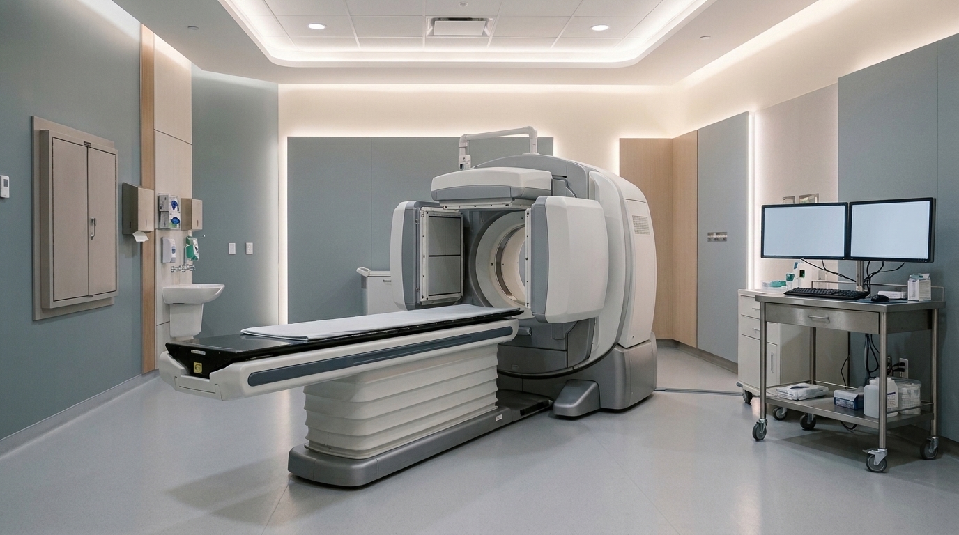
The pilot investigation evaluated a clinical cohort comprising five patients diagnosed with poorly differentiated thyroid carcinoma who collectively presented with eighteen metastatic bone lesions. As part of the pretherapeutic evaluation protocol, each participant received an administered diagnostic activity of 111 megabecquerels of iodine-131. Following administration, the investigators acquired serial quantitative single-photon emission computed tomography and computed tomography scans across eight discrete time points extending up to 120 hours post-injection.
Iodine-131 concentrations within blood and anatomical regions of interest were measured in units of megabecquerels per gram utilizing quantitative single-photon emission computed tomography and computed tomography technology. To validate the physical accuracy of these imaging-derived concentrations, blood iodine-131 activity concentrations determined from the volumetric imaging datasets were directly benchmarked against measurements obtained from concurrent physical blood samples using laboratory gamma counting. Statistical analysis confirmed an excellent level of agreement between the blood concentrations derived from quantitative single-photon emission computed tomography and computed tomography and those measured by conventional gamma counting, with the correlation reaching strong statistical significance at a p-value of less than 0.001.
Radiation absorbed dose rates, expressed in units of gray per hour, were estimated for circulating blood as well as metastatic tumour targets using advanced Monte Carlo computational simulations. These complex physical simulations were executed using a customized Geant4 simulation toolkit, and the computational modeling was independently cross-validated using the TOPAS simulation architecture. Cumulative radiation absorbed doses delivered to tumour tissues were subsequently calculated across the multi-timepoint surveillance interval by applying trapezoidal integration to the resulting absorbed dose rate trajectories over time. Following this pretherapeutic dosimetric mapping, clinical outcomes were assessed after the patients proceeded to receive subsequent high-dose radioactive iodine therapy.
Comparative dosimetric analysis revealed that blood absorbed doses calculated per unit of administered activity were comparable between the quantitative single-photon emission computed tomography and computed tomography approach and conventional blood-based calculation methods. A statistical comparison between the two dosimetry methodologies yielded a p-value of 0.6250, indicating no statistically significant difference in blood absorbed dose estimation.
When examining tumour targets, the measured iodine-131 concentration demonstrated a remarkably strong linear relationship with the simulated absorbed dose rate across the eighteen evaluated bone lesions. Linear regression analysis generated a predictive equation defined as tumour absorbed dose rate in gray per hour equal to 0.1256 multiplied by the tumour radioactive iodine concentration in megabecquerels per gram. This mathematical model demonstrated exceptional goodness-of-fit, characterized by a coefficient of determination R-squared value of 0.9918 and statistical significance at a p-value of less than 0.0001, providing a direct mathematical link between measured radioiodine concentration and tissue absorbed dose rate.
The research team evaluated dosimetric calculations when reducing the serial imaging protocol from the original eight time points down to four distinct imaging sessions.
In exploratory clinical outcome evaluations, bone metastases that achieved expected absorbed radiation doses exceeding 50 gray showed no disease progression following treatment. In contrast, metastatic lesions that received absorbed doses below the 50 gray threshold progressed far more frequently during follow-up, exhibiting an observed progression rate of 87.5 percent, a difference in therapeutic response that was statistically significant with a p-value of 0.0161.
The authors concluded that pretherapeutic quantitative iodine-131 single-photon emission computed tomography and computed tomography reliably determines blood and tumour dosimetry in poorly differentiated thyroid carcinoma patients presenting with bone metastases. Although the investigators emphasized that these findings remain preliminary and exploratory because of the small pilot cohort of five patients and eighteen lesions, the results support the clinical potential of the imaging platform as a practical tool for personalized radioactive iodine treatment planning. The researchers noted that larger prospective clinical studies are warranted to further evaluate and validate these clinical dose-response thresholds in expanded patient populations.
Subject of Research: Pretherapeutic quantitative I-131 SPECT/CT dosimetry for thyroid cancer bone metastases
Article Title: Pretherapeutic quantitative I-131 SPECT/CT for blood and tumour dosimetry in thyroid cancer patients with bone metastases: a pilot feasibility study
Article References: Kwon, K., Oh, D., Kim, J. H., Yoo, J., Song, Y. S., Sung, W., & Lee, W. W. (2026). Pretherapeutic quantitative I-131 SPECT/CT for blood and tumour dosimetry in thyroid cancer patients with bone metastases: a pilot feasibility study. EJNMMI Physics. https://doi.org/10.1186/s40658-026-00927-8
Image Credits: AI Generated
DOI: 10.1186/s40658-026-00927-8
Keywords: I-131, SPECT/CT, dosimetry, thyroid cancer, bone metastases, poorly differentiated thyroid carcinoma, radioactive iodine, nuclear medicine, Pretherapeutic, quantitative, SPECT, blood
News Source: Nathaniel Bowman. (November 7, 2026). Pilot Study Validates Quantitative SPECT/CT Dosimetry for Metastatic Thyroid Cancer. Scienmag.