


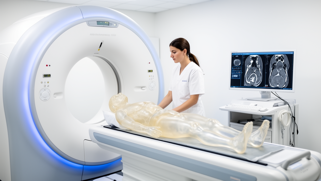
Peritoneal metastases, the spread of colorectal and other cancers to the lining of the abdominal cavity, are among the hardest lesions for radiologists to find on a routine CT scan. They are small, they sit in fat and along membranes, and conventional scanners simply do not give them enough contrast against the surrounding tissue. A team of French medical physicists and radiologists has now shown, in a carefully controlled phantom study, that a new kind of scanner can do measurably better, provided the images are reconstructed with the right settings.
The study, published in European Radiology Experimental, compared two imaging platforms built by Philips Healthcare: a Spectral CT7500 dual-layer CT system that uses conventional energy-integrating detectors, and an advanced research prototype spectral photon-counting CT, or SPCCT, equipped with cadmium-zinc-telluride photon-counting detectors. The researchers scanned an elliptical Multi-Energy CT phantom from Gammex and Sun Nuclear, fitted with sixteen removable inserts embedded in a water-equivalent background, and then systematically varied the reconstruction parameters on the photon-counting machine to see which combination produced the best images for detecting simulated peritoneal metastases.
The physics behind the comparison matters. Energy-integrating detectors, which are used in virtually all clinical CT scanners today, register the total energy deposited by many photons at once, and in doing so they blur away information about individual photon energies and add electronic noise to the signal. Photon-counting detectors instead register each X-ray photon individually and sort them by energy into multiple bins. That finer spectral sampling, the researchers note, allows more accurate characterization of photoelectric and Compton interactions and yields higher-quality spectral images, especially the low-energy virtual monoenergetic images, or VMIs, that radiologists use to boost the contrast of poorly enhancing lesions.
Both scanners acquired images at a tube voltage of 120 kVp, a rotation time of 0.5 seconds, a pitch factor of 1.241, and a fixed tube current of 273 mA, with the physical beam collimation set at 128 by 0.625 mm for the dual-energy system and 64 by 0.275 mm for the photon-counting prototype. The volume CT dose index was held close to 8.5 mGy on both systems, so the comparison was made at essentially equal radiation dose. From these acquisitions the team reconstructed virtual monoenergetic images at 40 keV, the energy level that a previous clinical study had shown improves the contrast-to-noise ratio of peritoneal metastases, particularly the non-mucinous type.
On the dual-energy scanner, the 40 keV images were reconstructed with level 4 of the hybrid iterative Spectral algorithm and the standard soft-tissue kernel B, settings that correspond to routine clinical practice for abdominal and pelvic spectral imaging at the investigators’ institutions. On the photon-counting prototype, the same 40 keV images were reconstructed with four levels of the iDose4 iterative reconstruction algorithm, spanning levels 5, 7, 9, and 11, crossed with two reconstruction kernels, STD-B and HRB, both recommended by the manufacturer for abdominal and pelvic spectral imaging. That produced eight distinct reconstruction combinations to evaluate against the clinical baseline.
The evaluation was task-based rather than a matter of visual impression. Using iQMetrix-CT software, the team computed the noise power spectrum from eight square regions of interest across 200 consecutive axial slices, quantifying both the magnitude of image noise and its texture through the average spatial frequency of the noise spectrum. Spatial resolution was measured through the task-based transfer function, calculated on an insert of blood-mimicking material with iodine at 2 mg/mL, whose theoretical contrast at 40 keV, 232 Hounsfield units, sits between the reported contrasts of non-mucinous and mucinous peritoneal metastases, 290 and 201 HU respectively. Detectability indices were then computed with a non-prewhitening observer model that includes an eye filter mimicking human visual sensitivity, using two simulated 5 mm lesions whose contrasts matched those two metastasis types.
The results showed a clear pattern. With the STD-B kernel, the photon-counting system produced lower noise magnitude, by 22.4 percent on average, and finer noise texture, with average noise-spectrum spatial frequencies 21.3 percent lower, while spatial resolution, measured as the frequency at which the transfer function falls to half its maximum, was 5.8 percent higher than with the HRB kernel at every iDose4 level. Detectability followed the noise: for both simulated lesion types, the detectability index was 22.3 percent higher with STD-B than with HRB. As expected, raising the iterative reconstruction level reduced noise on both kernels, with successive level increases cutting noise magnitude by roughly 29 percent each step.
Against the dual-energy baseline, the photon-counting system with the STD-B kernel matched or beat the clinical scanner on noise magnitude and detectability from level i7 upward, while the HRB kernel needed levels i9 and i11 to reach the same standing. Noise texture and spatial resolution were higher with the photon-counting system at nearly every setting, the single exception being spatial resolution at level i11 with the HRB kernel, which dipped slightly below the dual-energy value. Applying the study’s criterion that combinations yielding lower noise and higher texture, resolution, and detectability values count as optimal, the qualifying settings were levels i7, i9, and i11 with STD-B and levels i9 and i11 with HRB.
The overall winner was STD-B at iDose4 level 9, which the authors describe as the best compromise between noise texture, spatial resolution, and detectability for the simulated metastases. If the priority were image texture and spatial resolution alone, level i7 would win, though its noise and detectability only match the dual-energy scanner; if maximum detectability were the goal, level i11 would be the choice despite its softer texture and resolution. To check that these phantom findings meant something in a clinical setting, the team also scanned one patient, a 59-year-old woman with known non-mucinous peritoneal metastases, on both systems on the same day with matched contrast injection parameters and nearly identical dose, 8.2 versus 8.3 mGy. Two radiologists reviewing the images by consensus found that the photon-counting images showed better delineation of muscular fascia and better depiction of the parietal peritoneal nodule and omental fat stranding, with the STD-B kernel at level 9 again providing the best overall image quality.
The authors are careful about what this study does and does not establish. The work was performed on a single phantom whose inserts do not perfectly replicate real metastases, no statistical analysis was possible because each metric was calculated once per configuration, and the comparison relied on the reconstruction algorithms currently available for each system, meaning the observed gains cannot be attributed solely to the photon-counting detector technology itself. The patient example, they emphasize, is an in vivo demonstration rather than a clinical validation, and the findings must be confirmed in a larger cohort before standardized reconstruction protocols can be defined. Still, the direction of the result is unambiguous: at the same radiation dose, a properly tuned photon-counting CT can deliver less noise, finer noise texture, slightly better spatial resolution, and higher computed detectability of small, low-contrast peritoneal lesions than the dual-energy scanners in routine use today, a combination that could eventually sharpen the preoperative assessment of colorectal cancer patients whose treatment decisions hinge on seeing every peritoneal implant.
Subject of Research: Optimizing spectral photon-counting CT reconstruction parameters to improve detection of peritoneal metastases compared with energy-integrating detector dual-energy CT in a phantom study
Article Title: Optimization of spectral photon-counting CT reconstruction parameters for improved detection of peritoneal metastases compared with energy-integrating detector dual-energy CT: a phantom study
Article References: Greffier, J., Grange, R., Dabli, D., Houmeau, A., Antonuccio, M. N., Rousset, P., & Si-Mohamed, S. (2026). Optimization of spectral photon-counting CT reconstruction parameters for improved detection of peritoneal metastases compared with energy-integrating detector dual-energy CT: a phantom study. European Radiology Experimental, 10(1), Article 144. https://doi.org/10.1186/s41747-026-00816-w
Image Credits: AI Generated
DOI: 10.1186/s41747-026-00816-w
Keywords: photon-counting CT, dual-energy CT, peritoneal metastases, virtual monoenergetic images, image reconstruction, colorectal cancer, medical imaging, phantom study, detectability index, noise power spectrum, radiology, dose efficiency
News Source: Ophelia Keating. (October 30, 2026). Photon-counting CT beats dual-energy CT at spotting peritoneal metastases in phantom test. Scienmag.



