Credit: K Salem et al., University of Wisconsin School of Medicine and Public Health, Madison, WI
Physicians may soon have a new way to measure the efficacy or failure of hormone therapy for breast cancer patients, according to new research published in the February issue of The Journal of Nuclear Medicine. Researchers report that positron emission tomography (PET) imaging with 18F-fluorofuranylnorprogesterone (18F-FFNP) has been found to successfully measure changes in progesterone receptor (PR) levels resulting from a short-course estrogen treatment, also known as an estradiol challenge.
Estrogen-receptor (ER)-positive breast cancer is the most common class of breast cancer, affecting nearly 70 percent of patients. By participating in an estradiol challenge, physicians can determine the likelihood of potential benefit of hormonal therapies targeting ER for individual patients. Many hormone therapies interfere with the ability of estrogen to regulate the expression of PR protein, which is more pronounced in the presence of estrogen. As such, several PET tracers have been developed to monitor and analyze changes in the PR level during therapy. “Typically, anatomic size and proliferation biomarkers are analyzed to determine endocrine sensitivity,” said Amy M. Fowler, MD, PhD, assistant professor, Section of Breast Imaging, Department of Radiology, University of Wisconsin-Madison. “However, non-invasive detection of changes in PR expression with 18F-FFNP during an estradiol challenge may be an earlier indicator of the effectiveness of a specific hormone therapy.”
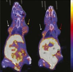
In this study, T47D human breast cancer cells (cells with estrogen and progesterone receptors, but without human epidermal growth factor receptor-2) and mice bearing T47D tumor xenografts were treated with estrogen to increase PR expression. The cells and mice were imaged with 18F-FFNP, and assays were conducted for cell uptake and tissue biodistribution. To investigate the separate role of PR-A and PR-B isoforms on overall 18F-FFNP binding, triple-negative MDA-MB-231 breast cancer cells were engineered to express either PR-A or PR-B. In vitro 18F-FFNP binding was measured by saturation and competitive binding assays, while in vivo uptake was measured with PET imaging.
In T47D cells treated with estrogen, an increase in 18F-FFNP uptake was measured at 48 hours after treatment; in mice with T47D tumor xenografts, increased uptake was seen at 48 and 72 hours after treatment. This increase in 18F-FFNP uptake also correlated with an increase in PR protein expression and proliferation. Results showed that there was no significant preferential 18F-FFNP binding or uptake by PR-A versus PR-B in PR isoform cell lines or tumor xenografts. “This is an important finding given the variability of PR isoform expression observed in breast cancer patients,” stated Fowler.
She continued, “Validation of PR imaging as a biomarker of endocrine sensitivity in patients before and after estradiol challenge could provide new opportunities in the field of molecular imaging and nuclear medicine for breast cancer imaging. Improved methods for testing endocrine sensitivity in patients could better inform decisions for optimal individualized ER-positive breast cancer therapy, potentially reducing morbidity and mortality.”
###
The authors of “Sensitivity and Isoform Specificity of 18F-Fluorofuranylnorprogesterone for Measuring Progesterone Receptor Protein Response to Estradiol Challenge in Breast Cancer” include Kelley Salem, Manoj Kumar, Kyle C. Kloepping, Ciara J. Michel and Ginny L. Powers, Department of Radiology, University of Wisconsin School of Medicine and Public Health Madison, Wisconsin; Yongjun Yan, Department of Radiology, University of Wisconsin School of Medicine and Public Health Madison, Wisconsin, and Department of Medical Physics, University of Wisconsin School of Medicine and Public Health Madison, Wisconsin; Justin J. Jeffery, University of Wisconsin Carbone Cancer Center, Madison, Wisconsin; Aparna M. Mahajan, Department of Pathology and Laboratory Medicine, University of Wisconsin School of Medicine and Public Health Madison, Wisconsin; and Amy M. Fowler, Department of Radiology, University of Wisconsin School of Medicine and Public Health Madison, Wisconsin, Department of Medical Physics, University of Wisconsin School of Medicine and Public Health Madison, Wisconsin, and University of Wisconsin Carbone Cancer Center, Madison, Wisconsin.
This study was made available online in July 2018 ahead of final publication in print in February 2019.
For questions or to schedule an interview with the researchers, please contact Rebecca Maxey at (703) 652-6772 or [email protected]. Current and past issues of The Journal of Nuclear Medicine can be found online at http://jnm.
About the Society of Nuclear Medicine and Molecular Imaging
The Society of Nuclear Medicine and Molecular Imaging (SNMMI) is an international scientific and medical organization dedicated to advancing nuclear medicine and molecular imaging, vital elements of precision medicine that allow diagnosis and treatment to be tailored to individual patients in order to achieve the best possible outcomes.
SNMMI’s more than 17,000 members set the standard for molecular imaging and nuclear medicine practice by creating guidelines, sharing information through journals and meetings, and leading advocacy on key issues that affect molecular imaging and therapy research and practice. For more information, visit http://www.
Media Contact
Rebecca Maxey
[email protected]
703-652-6772
Original Source
https:/
Related Journal Article
http://dx.


