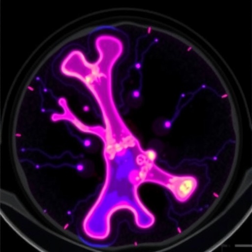
In a striking demonstration of how a tracer originally designed for the brain can illuminate disease far beyond the skull, physicians and nuclear medicine specialists at West China Hospital of Sichuan University have reported a case in which whole-body positron emission tomography with the amyloid-binding radiopharmaceutical 18F-florbetapir revealed widespread amyloid deposits throughout the skeleton of a patient with multiple myeloma complicated by systemic AL amyloidosis. The case, published in the European Journal of Nuclear Medicine and Molecular Imaging, was featured as an “Image of the Month,” a format reserved for visually and clinically compelling presentations that expand the diagnostic repertoire of practicing physicians. The images show multifocal, intense uptake of 18F-florbetapir within bones whose appearance on conventional imaging would otherwise have been attributed to the malignancy itself, distinguishing between two processes—plasma-cell infiltration and amyloid deposition—that often coexist but demand different therapeutic responses.
AL amyloidosis, the most common form of systemic amyloidosis, arises when a clone of plasma cells produces monoclonal immunoglobulin light chains that misfold into beta-pleated amyloid fibrils. These fibrils circulate in the blood and deposit in tissues, where they disrupt organ structure and function. The heart and kidneys are the classic targets, and cardiac involvement remains the principal determinant of prognosis. In patients whose amyloidosis is driven by an underlying myeloma, the diagnostic picture is especially tangled: bone pain, lytic lesions, and marrow abnormalities may reflect the proliferative plasma-cell disorder, the insidious accumulation of amyloid in bone, or both. Separating these entities in a living patient has long been a challenge, because amyloid deposits in bone marrow are difficult to sample and conventional imaging modalities cannot reliably distinguish amyloid infiltration from tumor replacement.
18F-florbetapir occupies a unique position in this evolving landscape. The tracer was developed and approved for the detection of beta-amyloid plaques in the brains of patients being evaluated for Alzheimer’s disease, where it binds with high affinity to aggregated amyloid. Because the fundamental pathology of all amyloidoses—beta-sheet fibrillar protein aggregation—is shared across amyloid types, researchers recognized more than a decade ago that florbetapir and related tracers could, in principle, flag amyloid anywhere in the body. Subsequent studies have borne this out. Work published in this same journal in 2018 demonstrated extracardiac uptake of 18F-florbetapir in patients with systemic amyloidosis, cheekily noting that the technique involved “more than hearts and minds,” and a 2019 study in the Journal of Nuclear Medicine showed that whole-body 18F-florbetapir PET/CT could detect multiorgan light-chain amyloidosis at early stages, well before functional impairment became evident. Comprehensive pathology analyses, including a 2022 study of nearly 1,500 bone marrow samples published in Amyloid, have established that bone marrow amyloid is far from rare, underscoring the relevance of skeletal imaging in these patients.
Against this backdrop, the West China Hospital case provides an unusually vivid illustration of what skeletal amyloid burden looks like on molecular imaging. The patient, who had established multiple myeloma with associated systemic AL amyloidosis, underwent whole-body PET/CT after intravenous administration of 18F-florbetapir. The resulting images, acquired roughly an hour after injection, revealed multiple foci of avid tracer accumulation distributed across the axial and appendicular skeleton. On the fused images, these hotspots stood out against a background of physiological tracer distribution, and their correspondence with lytic skeletal abnormalities on the CT component suggested that amyloid had deposited within or adjacent to bone lesions caused by the myeloma. The pattern—multifocal, skeletal, and anatomically widespread—documents a degree of osseous amyloid involvement that is rarely visualized so directly and carries immediate implications for how clinicians stage and monitor the disease.
The technical logic of the scan merits careful unpacking, because it explains why the images are so informative. Florbetapir contains an 18F-labeled stilbene derivative that crosses cell membranes and binds selectively to aggregated, fibrillar structures rich in cross-beta-sheet conformation, with minimal uptake in healthy tissue. When the radiolabeled molecule accumulates in a tissue, the positrons it emits annihilate with electrons to produce pairs of 511-keV photons detected in coincidence by the PET scanner, allowing reconstruction of a three-dimensional map of tracer concentration. Co-registration with computed tomography provides the anatomical scaffold onto which this molecular signal is projected, so that each focus of uptake can be localized to a specific vertebra, rib, or long bone. In this patient, the CT component showed myeloma-typical osteolytic destruction, while the PET component revealed that many of these sites were also repositories of amyloid fibrils—information that no conventional modality could have supplied.
The clinical significance of identifying skeletal amyloid deposits is twofold. First, it refines prognosis and symptom attribution. Amyloid deposition in bone and periarticular structures can contribute to bone pain, pathological fracture risk, and even carpal tunnel syndrome and other entrapment neuropathies in AL patients. Knowing that a patient’s skeleton is diffusely infiltrated by amyloid helps clinicians interpret symptoms that might otherwise be blamed on tumor progression, and it informs decisions about analgesia, orthopedic intervention, and the pacing of therapy. Second, and perhaps more importantly, molecular imaging of amyloid offers a potential surrogate marker for monitoring response. In AL amyloidosis, the goal of therapy is to suppress the light-chain-producing clone with regimens such as anti-CD38 antibodies, proteasome inhibitors, or autologous stem-cell transplantation, thereby halting new amyloid formation. Existing monitoring relies on serum free light-chain measurements and cardiac biomarkers, neither of which reveals where amyloid has already accumulated or whether established deposits are resolving. Serial 18F-florbetapir PET/CT, as this case suggests, could eventually complement biochemical monitoring by visualizing the anatomical distribution and temporal evolution of the amyloid burden itself.
The case also speaks to a broader diagnostic dilemma: differentiating AL amyloidosis from its rarer counterparts, particularly transthyretin amyloidosis and amyloid light-chain disease arising without an overt plasma-cell neoplasm. Bone scintigraphy with 99mTc-labeled phosphates is well established for identifying cardiac transthyretin amyloid, but it performs poorly for AL disease and provides no information about skeletal amyloid. Biopsy of an accessible organ—typically abdominal fat or bone marrow—remains the diagnostic gold standard, yet biopsy is invasive, subject to sampling error when deposits are patchy, and impossible to repeat casually for disease monitoring. A whole-body imaging technique that flags amyloid noninvasively, and that can be repeated over the course of therapy, therefore fills a genuine unmet need, especially in myeloma patients in whom marrow involvement is already suspected on other grounds.
The authors of the report, Xiao Zhong and Xiaohong Ou of the Nuclear Medicine Department at West China Hospital, emphasize that the images were obtained under standard clinical imaging protocols with appropriate ethical oversight; the study was approved by the hospital’s ethics committee and conducted in accordance with the Declaration of Helsinki, with informed consent obtained from the patient for both participation and publication of the images. The work was supported by the National Natural Science Foundation of China through a Youth Science Fund grant, reflecting growing institutional investment in molecular imaging applications for hematologic and amyloid diseases. The authors declare no competing interests, and the de-identified patient data underlying the report are available from the corresponding author upon reasonable request.
For the nuclear medicine community, the case adds to a growing repertoire of amyloid-PET applications that extend well beyond the tracer’s original neurological mandate. As whole-body PET scanners with improved sensitivity and spatial resolution become more widely available, the feasibility of surveying the entire skeleton for amyloid deposition increases accordingly. Long-wavelength total-body scanners, motion-corrected reconstruction algorithms, and quantitative uptake metrics such as standardized uptake values could eventually allow clinicians to grade skeletal amyloid burden numerically, track it longitudinally, and correlate it with clinical outcomes in a way that qualitative image inspection cannot. The present case, with its dramatic multifocal skeletal signal, offers a template for what such quantitative monitoring might target.
For patients with myeloma and suspected amyloidosis, the takeaway is more immediate: skeletal amyloid involvement, long an invisible companion to the malignancy, can now be seen. The images from Chengdu show that when a light-chain clone churns out misfolding proteins, the resulting fibrils may accumulate not only in the heart and kidneys but across the entire skeleton, insinuating themselves into the very bones the myeloma is destroying. Recognizing this dual pathology on a single scan transforms the diagnostic conversation, replacing inference with direct visualization and opening a path toward imaging-guided assessment of one of medicine’s most protean protein-misfolding diseases. As amyloid-targeted PET tracers move from Alzheimer’s clinics into the systemic arena, cases like this one signal that the era of seeing amyloid whole—organ by organ, bone by bone—has arrived.
Subject of Research: Multifocal skeletal amyloid deposits visualized by 18F-florbetapir PET/CT in a patient with multiple myeloma-associated systemic AL amyloidosis
Subject of Research: Medicine
Article Title: Multifocal skeletal 18F-florbetapir uptake on PET/CT in myeloma-associated systemic AL amyloidosis
Article References: Zhong, X., & Ou, X. (2026). Multifocal skeletal 18F-florbetapir uptake on PET/CT in myeloma-associated systemic AL amyloidosis. European Journal of Nuclear Medicine and Molecular Imaging. https://doi.org/10.1007/s00259-026-08156-y
Image Credits: AI Generated
DOI: 10.1007/s00259-026-08156-y
Keywords: 18F-florbetapir, PET/CT, AL amyloidosis, multiple myeloma, skeletal amyloid uptake, amyloid imaging, light-chain amyloidosis, nuclear medicine, whole-body PET, amyloid fibrils
Cite Scienmag News
APA MLA Chicago
Ophelia Keating. (September 5, 2026). PET/CT reveals bone uptake of amyloid tracer in AL amyloidosis. Scienmag. https://scienmag.com/pet-ct-reveals-bone-uptake-of-amyloid-tracer-in-al-amyloidosis/
Ophelia Keating. “PET/CT reveals bone uptake of amyloid tracer in AL amyloidosis.” Scienmag, 5 September 2026, https://scienmag.com/pet-ct-reveals-bone-uptake-of-amyloid-tracer-in-al-amyloidosis/. Accessed 5 September 2026.
Ophelia Keating. “PET/CT reveals bone uptake of amyloid tracer in AL amyloidosis.” Scienmag. September 5, 2026. https://scienmag.com/pet-ct-reveals-bone-uptake-of-amyloid-tracer-in-al-amyloidosis/
Copy citation Download RIS
Tags: 18F-florbetapir bone uptake18F-florbetapir in systemic amyloidosisadvanced imaging for amyloid localizationadvanced imaging for amyloidosisAL amyloidosis detectionAL amyloidosis diagnosisamyloid deposition in bonesamyloid imaging in multiple myelomaamyloid PET/CT case studyamyloid PET/CT imagingamyloid tracer in skeletal imagingamyloid tracer uptake in bonesbone amyloid depositiondifferential diagnosis of bone lesionsdifferentiation of plasma-cell infiltration and amyloid depositsnon-invasive diagnosis of systemic amyloidosisnuclear medicine in amyloidosisPET imaging in multiple myelomaPET/CT in bone amyloid detectionsystemic amyloid depositssystemic amyloidosis diagnostic imaging