In a retrospective single-center study evaluating noninvasive diagnostics for transthyretin amyloid cardiomyopathy, researchers found that patients exhibiting Perugini grade 3 tracer uptake on technetium-99m pyrophosphate scintigraphy presented with significantly more advanced structural remodeling, expanded myocardial extracellular volume, more frequent right ventricular involvement, and greater functional impairment than those with Perugini grade 2 uptake. Although current international diagnostic guidelines classify both visual Perugini grades 2 and 3 as positive diagnostic criteria for transthyretin amyloid cardiomyopathy once clonal light chain amyloidosis has been ruled out, comparative phenotypic analyses between these two positive scintigraphic tiers have remained limited. The seven-year investigation, conducted at Lithuanian University of Health Sciences Hospital Kauno Klinikos and published in the Journal of Cardiovascular Imaging, demonstrates that higher scintigraphic uptake is accompanied by measurable differences across multimodality imaging markers, circulating cardiac biomarkers, and physical functional capacity within a specialized clinical referral population.
Transthyretin amyloid cardiomyopathy has gained increasing recognition as a major underlying cause of heart failure with preserved ejection fraction, especially following the development and clinical validation of noninvasive nuclear imaging pathways. Historically, definitive identification of the condition required an invasive endomyocardial biopsy, an intervention whose associated procedural hazards and restricted technical availability frequently outweighed prospective clinical benefits when targeted, disease-modifying therapies did not yet exist. The validation of bone-avid radiotracer scintigraphy alongside the commercial introduction and therapeutic adoption of targeted transthyretin-modifying treatments has fundamentally redirected clinical practice toward the detection of earlier disease signs. In nuclear imaging protocols, visual Perugini grading compares the relative intensity of myocardial radiotracer accumulation against uptake visible within the adjacent bony ribcage, serving as the central noninvasive metric for diagnostic confirmation.
The retrospective review encompassed all 302 technetium-99m pyrophosphate scintigraphy examinations performed for clinically suspected cardiac amyloidosis at Kauno Klinikos between 2018 and 2025. Planar imaging identified cardiac-region radiotracer accumulation in 103 scans, representing 34.1 percent of the total volume evaluated. Grade 1 visual uptake accounted for 48 scans, or 46.6 percent of those with reported activity, while grade 2 uptake was observed in 10 scans and grade 3 uptake was recorded in 45 scans. Importantly, none of the patients categorized with Perugini grade 1 uptake received a subsequent diagnosis of transthyretin amyloid cardiomyopathy throughout a median follow-up period of 28.0 months. Hybrid single-photon emission computed tomography combined with computed tomography, available for 27 of the grade 1 cases, revealed that true myocardial uptake occurred in only three individuals, whereas the remaining 24 cases reflected residual blood-pool tracer retention that planar views had misidentified as myocardial retention, reinforcing clinical guideline recommendations requiring tomographic confirmation.
The definitive study cohort comprised 53 patients with confirmed transthyretin amyloid cardiomyopathy following the diagnostic exclusion of two patients with grade 2 uptake who were determined to have amyloid light chain amyloidosis. Within this confirmed cohort of 53 individuals, eight patients, or 15.1 percent, presented with Perugini grade 2 uptake, whereas 45 patients, or 84.9 percent, exhibited Perugini grade 3 uptake. Genetic sequencing was executed across every diagnosed patient, identifying variant-type transthyretin amyloid cardiomyopathy in two individuals and wild-type disease in the remaining 51 participants. Reflecting evolving clinical awareness and protocol adoption, annual diagnostic identification increased considerably across the observation window, with 40 of the 53 cohort cases diagnosed during the 2023 to 2025 period alone. Noninvasive diagnostic confirmation accounted for 45 of the 53 total cases, rising to represent over 80 percent of annual institutional diagnoses from 2023 onward.
Comparative clinical assessments between the scintigraphic groups revealed substantial differences in physical performance and functional limitation despite comparable chronological age and demographic distribution. The average patient age at diagnosis was 78 years across the cohort, with no meaningful divergence between the grade 2 group at 77.8 years and the grade 3 group at 78.2 years, and men constituted 69.8 percent of the overall population. However, patients with grade 3 uptake demonstrated significantly worse New York Heart Association functional class and achieved shorter distances during standardized six-minute walk testing, averaging 298 meters compared to 366 meters recorded in grade 2 patients. Furthermore, permanent cardiac pacemaker implantation had been performed exclusively in grade 3 individuals, occurring in 22.2 percent of that group.
Patients with grade 3 uptake exhibited significantly higher maximal wall thickness, averaging 16.9 millimeters compared to 14.6 millimeters in grade 2 individuals. Left ventricular systolic function was also more impaired in the grade 3 tier, evidenced by a lower left ventricular ejection fraction of 49.1 percent versus 53.8 percent and a worse absolute global longitudinal strain of 12.5 percent compared to 16.0 percent. Furthermore, the classical echocardiographic pattern of apical sparing occurred with significantly greater frequency among grade 3 patients, appearing in 88.9 percent of scans compared to 50.0 percent among grade 2 patients, whereas left atrial volume index measurements remained statistically comparable between groups.
Multiparametric cardiac magnetic resonance imaging, completed within four months of scintigraphy in a subset of 38 patients consisting of eight grade 2 cases and 30 grade 3 cases on a 3.0-Tesla scanner, further corroborated the presence of greater myocardial infiltration. Left ventricular mass index was significantly elevated among grade 3 patients, reaching 106.0 grams per square meter compared to 68.1 grams per square meter in grade 2 patients. Extracellular volume fraction, determined from hematocrit-corrected pre- and post-contrast modified Look-Locker inversion recovery T1 mapping sequences across basal and mid-ventricular septal myocardium, was markedly higher in grade 3 disease, measuring 45.2 percent versus 35.1 percent. Right ventricular structural involvement was also more pronounced in grade 3 patients, who exhibited thicker right ventricular free walls measuring 6.2 millimeters versus 4.0 millimeters and a higher frequency of right ventricular late gadolinium enhancement, which was documented in 93.3 percent of grade 3 scans compared to 37.5 percent of grade 2 scans.
Analysis of circulating cardiac biomarkers highlighted distinct biochemical profiles that reflected divergent underlying pathophysiological mechanisms. Patients exhibiting Perugini grade 3 uptake had markedly higher circulating levels of cardiac troponin I, averaging 0.099 micrograms per liter compared to 0.025 micrograms per liter observed in the grade 2 cohort. In contrast, concentrations of brain natriuretic peptide did not show a statistically significant difference between the two scintigraphic groups, registering at 332 nanograms per liter in grade 3 versus 139 nanograms per liter in grade 2. Because troponin I and brain natriuretic peptide showed no mutual correlation within the cohort, the authors noted that troponin release likely captures ongoing microvascular compromise, direct cellular cytotoxicity, and active amyloid infiltration, whereas natriuretic peptide elevations primarily reflect hemodynamic wall stress and filling chamber pressures secondary to myocardial stiffening.
Rank-based correlation testing demonstrated robust associations linking visual Perugini grades to structural, tissue, and biochemical markers of disease severity. Perugini grade exhibited significant positive rank correlations with cardiac magnetic resonance left ventricular mass index, yielding a correlation coefficient of 0.608, and with extracellular volume fraction at 0.530. Visual grade also correlated significantly with circulating troponin I at 0.549 and brain natriuretic peptide at 0.355. In secondary evaluations, brain natriuretic peptide correlated positively with echocardiographic maximal wall thickness, left ventricular mass index, and extracellular volume fraction, while inversely correlating with absolute global longitudinal strain. Circulating troponin I correlated significantly with extracellular volume fraction, global longitudinal strain, and right ventricular ejection fraction, but exhibited no statistically significant relationship with myocardial mass measures or functional walk distances.
The study authors emphasized several structural limitations inherent to their single-center, retrospective analysis, particularly the modest size of the grade 2 comparator subgroup and the lack of long-term survival outcomes. Perugini scoring was conducted by a single experienced nuclear medicine reader without intraobserver or interobserver reproducibility assessments, introducing potential visual grading variability. Magnetic resonance examinations were unavailable in 15 diagnosed patients due to contraindications, scanner availability, or image quality, and N-terminal pro-brain natriuretic peptide was not measured, preventing the application of standard clinical staging algorithms. Consequently, the authors explicitly cautioned that their observations reflect cross-sectional phenotypic differences in a referral population and must not be interpreted as evidence that visual Perugini grades provide independent prognostic risk stratification.
Subject of Research: Multimodality imaging and phenotypic characterization of Perugini scintigraphic grades in transthyretin amyloid cardiomyopathy
Article Title: Multimodality imaging characterization of Perugini scintigraphic grades in transthyretin amyloid cardiomyopathy: a single-center experience
Article References: Bucius, P., Zarambaite, E., Lusaite, K., Streckis, M., Karuzas, A., Plisiene, J., Sakalyte, G., Lapinskas, T., Vajauskas, D., Jankauskas, A., & Ereminiene, E. (2026). Multimodality imaging characterization of Perugini scintigraphic grades in transthyretin amyloid cardiomyopathy: a single-center experience. Journal of Cardiovascular Imaging, 34(1), Article 15. https://doi.org/10.1186/s44348-026-00075-8
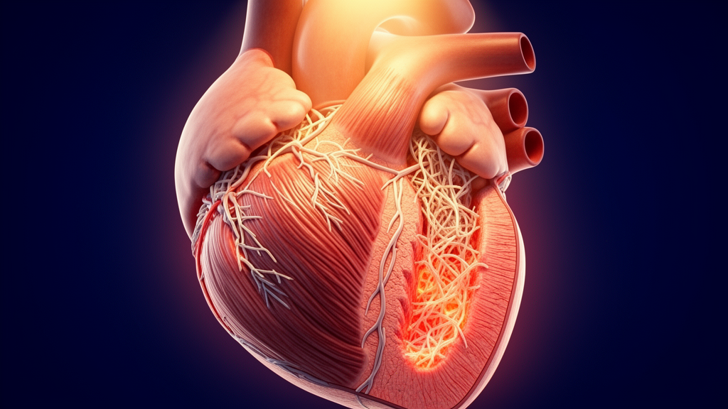
Image Credits: AI Generated
DOI: 10.1186/s44348-026-00075-8
Keywords: transthyretin amyloid cardiomyopathy, ATTR-CM, Perugini grading, technetium-99m pyrophosphate, 99mTc-PYP scintigraphy, cardiac magnetic resonance, echocardiography, extracellular volume fraction, troponin I, brain natriuretic peptide, SPECT-CT imaging, left ventricular mass index, global longitudinal strain, myocardial amyloidosis, right ventricular involvement, noninvasive cardiac diagnostics
News Source: Ophelia Keating. (November 19, 2026). Perugini Grade Three Scintigraphy Marks Advanced Cardiac Amyloidosis Structural Remodeling. Scienmag.