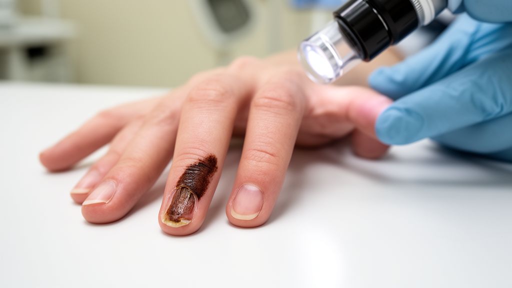
A persistent nail problem that refuses to heal deserves more attention than most people give it. A case report published in the journal Skin describes a 74-year-old Chinese man whose left thumbnail slowly detached from the nail bed over the course of a year, a condition known as distal onycholysis. What looked like an ordinary fungal infection turned out to be something far more dangerous: subungual amelanotic melanoma, a rare and pigment-free form of nail cancer that is notoriously easy to miss.
The patient had no family history of melanoma and no history of trauma to the finger, two factors that might otherwise have pointed clinicians toward a simpler explanation. In the early stages, the lesion was misdiagnosed as onychomycosis, the medical term for a fungal nail infection, at a nail salon where he received repeated manicure treatments. Unsurprisingly, these cosmetic interventions produced no improvement. When physicians eventually debrided the detached nail plate, they found pale-red proliferative tissue with erosion underneath, a finding that prompted a biopsy of the nail bed.
Histopathological examination revealed atypical melanocytes with marked cellular atypia infiltrating both the epidermis and the dermis. Immunohistochemical staining confirmed the diagnosis: the tumor cells tested positive for Melan-A, HMB45, and Prame, three markers commonly used to identify melanocytic malignancies, and the Ki-67 proliferation index was high, indicating rapid cell division. The final diagnosis was subungual amelanotic melanoma, a variant that lacks the dark pigmentation typically associated with melanoma and therefore mimics a range of benign conditions.
Staging and treatment followed established oncological protocols. Imaging studies found no evidence of metastasis, and the patient underwent wide local excision of the tumor along with a sentinel lymph node biopsy, which came back negative. He was classified as AJCC IIIC, designated T3bN1cM0, reflecting a tumor of substantial local extent with regional lymph node involvement but no distant spread. Adjuvant therapy combined interferon α1b, an immune-stimulating cytokine, with toripalimab, a checkpoint inhibitor antibody that blocks the PD-1 pathway and enhances the immune system’s ability to recognize tumor cells. At six months of follow-up, there was no local recurrence and no distant metastasis, an encouraging outcome given the aggressive staging.
The epidemiology of subungual melanoma varies dramatically across populations. In Asian and African populations, subungual tumors account for roughly 10 to 25 percent of all melanomas, whereas in Caucasian populations the figure drops to between 1 and 3 percent. This disparity is thought to relate to differences in pigmentation of the nail apparatus and surrounding skin, although the exact mechanisms remain an active area of research. The condition typically presents as pigmented streaks or masses beneath or within the nail plate, patterns that dermatologists are trained to recognize as potential warning signs.
The amelanotic variant, however, lacks pigment entirely, which strips clinicians of the most obvious visual clue. As a result, these tumors are frequently mistaken for fungal infections, viral warts, pyogenic granuloma, a benign but rapidly growing vascular lesion, or squamous cell carcinoma. Each of these misdiagnoses can delay definitive treatment by months or even years, and in melanoma, delay can be fatal. The case report emphasizes that persistent, isolated onycholysis or any non-pigmented lesion beneath the nail should prompt histopathological evaluation without delay, meaning a biopsy should be performed to obtain tissue for microscopic examination.
From a technical standpoint, the diagnostic pathway in this case illustrates the value of combining clinical observation with laboratory confirmation. The pale-red proliferative tissue visible after debridement was a subtle finding, one that could easily have been dismissed as granulation tissue from chronic irritation. Only the biopsy, followed by careful histopathological and immunohistochemical analysis, revealed the true nature of the lesion. The positivity for Melan-A and HMB45, both markers of melanocytic differentiation, together with Prame, a marker associated with malignant melanoma, and the elevated Ki-67 index, which measures the fraction of cells actively dividing, collectively ruled out benign mimics and confirmed a malignant process.
The surgical management also demonstrates current best practice for localized melanoma. Wide local excision aims to remove the tumor with a margin of healthy tissue, reducing the risk of local recurrence, while sentinel lymph node biopsy samples the first lymph node in the drainage pathway to determine whether cancer cells have begun to spread. A negative sentinel node biopsy, as in this case, is a favorable prognostic indicator. The addition of adjuvant therapy with interferon α1b and toripalimab reflects a modern approach to managing higher-risk disease, leveraging both immune stimulation and checkpoint blockade to reduce the likelihood of recurrence.
The broader lesson for the public is straightforward but important. A nail that separates from the nail bed and does not heal, particularly when it affects only one digit and there is no history of trauma or fungal infection, warrants medical evaluation rather than repeated cosmetic treatment. Nail salons are not diagnostic facilities, and manicures cannot cure an underlying malignancy. Similarly, any new growth, erosion, or discoloration under a nail, whether pigmented or not, should be examined by a healthcare professional capable of performing a biopsy. The work, titled Subungual Amelanotic Melanoma with Onycholysis as the First Manifestation, was published in Skin on August 21, 2026, and serves as a reminder that even the most mundane-seeming nail complaints can occasionally conceal a serious and life-threatening disease.
Subject of Research: Subungual amelanotic melanoma presenting as persistent nail separation
Article Title: Persistent nail separation may hide a rare melanoma
Article References: Persistent nail separation may hide a rare melanoma. Original publication (Bibliographic details unavailable.)
Image Credits: AI Generated
DOI: Not provided
Keywords: subungual melanoma, amelanotic melanoma, onycholysis, nail cancer, misdiagnosis, biopsy, immunohistochemistry, toripalimab, wide local excision, dermatology, Persistent, nail
News Source: Nathaniel Bowman. (October 19, 2026). Persistent nail separation may hide a rare melanoma. Scienmag.