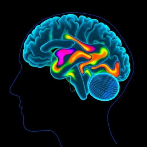
In a groundbreaking development that could revolutionize the early diagnosis and personalized treatment of Parkinson’s disease, researchers have demonstrated that individual cases of this neurodegenerative disorder can be robustly classified using magnetoencephalography (MEG). This advanced neuroimaging technique, which measures magnetic fields produced by neural activity, offers unprecedented insight into the brain’s dynamic functional architecture affected by Parkinson’s. The study, conducted by Roberts, Hardy, Pan, and colleagues and published in npj Parkinson’s Disease in 2026, marks a significant leap forward in the potential for objective and precise biomarker-based diagnostics in neurodegenerative diseases.
Parkinson’s disease, characterized by motor symptoms such as tremors, rigidity, and bradykinesia, is notorious for its clinical heterogeneity and overlapping symptomatology with other neurological conditions. Traditional diagnostic approaches largely rely on clinical observation and response to dopaminergic therapies, which can lead to misdiagnosis and delayed interventions. The promise of MEG lies in its ability to non-invasively capture neural oscillations and network dysfunctions at millisecond temporal resolution, facilitating a more nuanced understanding of Parkinson’s pathophysiology.
The research team employed sophisticated machine learning algorithms to analyze MEG data collected from patients diagnosed with Parkinson’s disease and healthy control subjects. By focusing on frequency-specific patterns of neural oscillations, cortical connectivity disruptions, and spectral power alterations in resting-state brain activity, the investigators were able to develop robust classifiers capable of differentiating individual Parkinson’s cases with remarkable accuracy. This classification was achieved without reliance on clinical symptoms alone, underscoring the power of MEG to uncover subtle neurophysiological signatures unique to the disease.
One particularly compelling aspect of this study is the ability of MEG to detect pathophysiological changes in early or prodromal stages of Parkinson’s disease. By identifying unique MEG biomarkers that correlate with disease severity and progression, clinicians may be equipped to initiate treatment regimens earlier, potentially slowing neurodegeneration. Moreover, this approach could facilitate patient stratification in clinical trials, ensuring that therapies are targeted to those most likely to benefit based on their individual brain activity profiles.
The analysis revealed that abnormal beta-band oscillations—widely implicated in motor dysfunction in Parkinson’s—alongside alterations in gamma and theta rhythm dynamics, serve as key discriminants between affected and unaffected brains. These oscillatory abnormalities reflect disrupted communication within basal ganglia-thalamo-cortical circuits, which are central to motor control. The high spatial and temporal resolution of MEG allowed researchers to pinpoint these disruptions with precision, further advancing our understanding of Parkinsonian neural circuitry.
Importantly, the research highlighted the reproducibility and generalizability of MEG-based classification across different subjects and scanning sessions. This robustness is critical for clinical application, where consistency in biomarker detection underpins reliable diagnosis and ongoing monitoring. The study’s findings suggest that MEG, when combined with advanced computational models, could serve as a frontline tool in the neurological clinic, augmenting traditional assessment paradigms.
The researchers also addressed potential confounding factors such as medication status, patient age, and comorbidities, ensuring that the MEG markers identified are intrinsic to Parkinson’s pathology rather than external influences. Their rigorous methodological framework strengthens confidence in the interpretability and utility of the results.
Beyond diagnosis, the implications of this work extend into therapeutic innovation. Understanding individualized neural signatures offers a path toward precision neuromodulation strategies, such as tailored deep brain stimulation protocols that adapt to the patient’s distinct oscillatory patterns. As MEG technology becomes more accessible and integrated with real-time analytics, such dynamic modulation could transform the management of Parkinson’s symptoms.
While MEG systems have traditionally been costly and limited to specialized research centers, advances in sensor technology and data processing are rapidly democratizing access to this powerful modality. Portable and wearable MEG devices on the horizon promise to bring this diagnostic capability closer to routine clinical settings, enabling longitudinal monitoring and early identification of disease shifts.
This study’s success also sets a precedent for applying MEG classification techniques to other neurodegenerative disorders, such as Alzheimer’s disease and multiple system atrophy, where clinical overlap complicates diagnosis. The precision and speed of MEG open new avenues for disentangling complex brain pathologies through direct measurement of their electrophysiological fingerprints.
Undoubtedly, challenges remain in validating MEG-based classifiers across larger and more diverse populations, and integrating this approach with multimodal biomarkers—such as molecular imaging and cerebrospinal fluid analyses—will be essential to capture the full spectrum of disease biology. Nevertheless, the robust classification of Parkinson’s disease on an individual level represents a landmark achievement in neurology and biomedical engineering.
As neuroscience advances into the era of personalized medicine, tools like MEG that marry non-invasive recording with sophisticated computational analysis will play a transformative role. The work by Roberts and colleagues not only enhances diagnostic precision but also enriches our mechanistic understanding of Parkinson’s, paving the way for targeted interventions tailored to the unique neurophysiological profile of each patient.
Future research will focus on longitudinal studies to track disease progression through MEG biomarkers, explore their predictive power in preclinical populations, and refine integration with therapeutic monitoring. By harnessing the intricate ballet of brain rhythms, this modality stands to elevate both clinical care and research in neurodegenerative disorders.
In summary, the successful classification of individual Parkinson’s cases via magnetoencephalography exemplifies the power of merging advanced neurotechnology with machine learning. This synergy promises to shift the paradigm from symptomatic diagnosis to direct neural characterization, offering hope for earlier, more accurate detection and more effective individualized treatments for Parkinson’s disease and beyond.
Subject of Research: Classification and diagnosis of Parkinson’s disease using magnetoencephalography (MEG)
Article Title: Individual cases of Parkinson’s disease can be robustly classified using magnetoencephalography
Article References:
Roberts, G., Hardy, S., Pan, Y. et al. Individual cases of Parkinson’s disease can be robustly classified using magnetoencephalography. npj Parkinsons Dis. (2026). https://doi.org/10.1038/s41531-026-01345-4
Image Credits: AI Generated
Tags: advanced neuroimaging techniques in neurologybiomarker-based diagnostics for Parkinson’scortical connectivity disruptions in Parkinson’searly detection of Parkinson’s diseasefrequency-specific neural activity analysisfunctional brain architecture in Parkinson’smachine learning for neurodegenerative disease classificationMEG neuroimaging in Parkinson’sneural oscillations in Parkinson’s diseaseneurodegenerative disease machine learning modelsParkinson’s disease diagnosis with magnetoencephalographypersonalized treatment for Parkinson’s


