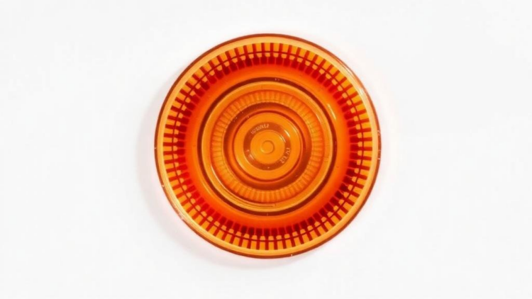
Cancer is projected to affect more than 30 million people worldwide by 2050, a staggering 75 percent increase over 2022 figures, with deaths rising by roughly 90 percent. As the global burden grows, the race to detect tumors earlier and monitor them more precisely has become one of the most urgent challenges in medicine. A study published in Bioengineering & Translational Medicine now reports a deceptively simple solution: a palm-sized plastic chip shaped like a flattened spiral that can pull rare tumor cells out of a blood sample in less than half an hour, without the harsh pre-treatment steps that plague conventional methods.
The device, developed by Isidora Panez-Toro and colleagues, targets circulating tumor cells, or CTCs, the wandering seeds of metastasis that shed from primary tumors into the bloodstream. These cells are extraordinarily scarce, often fewer than ten per ten milliliters of blood, and they survive in circulation for less than an hour before immune attack and shear stress destroy them. Catching them has long been compared to finding a handful of specific needles in a haystack the size of a stadium. Yet their capture matters enormously, because CTCs carry information about tumor biology that can guide therapy selection and track how a cancer evolves in real time.
The team’s approach rests on a physical phenomenon first described in 1961, when Segré and Silberberg observed that particles flowing through a straight channel migrate, seemingly spontaneously, to orderly lateral positions. Scientists later worked out the forces behind this inertial focusing: a shear-induced lift that pushes particles away from the channel’s centerline and a wall-induced lift that repels them from the walls. When the channel is coiled into a spiral, a secondary rotating flow known as Dean flow adds a drag force that nudges particles sideways. The delicate balance between these lift forces and Dean drag depends on particle size, which means that cells of different dimensions settle into different equilibrium positions and can be steered into separate outlets, purely by fluid dynamics, with no labels, magnets, or lasers.
What distinguishes the new work is its geometry. The researchers built two devices sharing identical inlets, channel cross-sections, and outlet architecture, but with different spiral shapes: a classic Archimedean, or circular, spiral and an oval one. The oval design introduces an oscillation in the radius of curvature with each loop, which modulates the secondary Dean forces and, the authors hypothesized, could sharpen the redistribution of cells and particles. Both chips feature six counterclockwise loops, a rectangular channel 530 micrometers wide and 140 micrometers tall, and three outlets at the periphery. The round spiral’s channel runs 21 centimeters; the oval’s stretches to 25 centimeters, with a short radius of 6.5 millimeters and a long radius of 11 millimeters.
To model blood, the team used fluorescent polystyrene beads of three sizes: 10 micrometers to mimic platelets and red blood cells, 13 micrometers to stand in for white blood cells, and 26 micrometers to represent tumor cells. The beads were suspended in bovine serum albumin solutions chosen to reproduce the density and viscosity of whole blood, which is a non-Newtonian fluid, and the devices were tested under both symmetric flow, with two syringes each delivering 800 microliters per minute, and asymmetric flow, with a fast buffer stream of 2800 microliters per minute meeting a slower particle stream of 600 microliters per minute. Under asymmetric conditions, the round spiral concentrated 13-micrometer beads at the middle outlet with about 90 percent relative isolation, while 26-micrometer beads went to the inner outlet, and the oval chip achieved inner-outlet isolation above 97 percent for the large beads.
The researchers then diluted the carrier fluid, dropping the albumin concentration from 30 percent to 16, 8, and 4 percent to simulate the blood dilution common in other protocols. The results were telling: lower density degraded the focusing of mid-sized particles and, in the oval device, split their distribution between outlets, while large particles remained robustly focused at the inner outlet. In other words, concentrating the sample rather than diluting it preserves the hydrodynamic precision, a finding that runs counter to the widespread practice of diluting blood tenfold or more before microfluidic processing, a step that inflates processing times to hours and risks losing precious cells.
Real cells, however, are not rigid beads. When the team flowed GFP-expressing MNNG-HOS osteosarcoma cells, which average about 17 micrometers in diameter, through the round spiral, the cells behaved like the 13-micrometer particles rather than the 26-micrometer ones, likely because cellular deformability lets them squeeze under hydraulic forces that would deflect a solid bead. The picture grew more complex in whole blood spiked with tumor cells: under asymmetric flow, cells scattered between the middle and outer outlets, an inversion the authors attribute to the sheer number of blood cells overwhelming the equilibrium forces. Switching to symmetric flow restored order, and the oval spiral proved superior, directing 38,026 of 40,000 spiked cells to the inner outlet, compared with 31,657 for the round design.
The decisive breakthrough came from narrowing the channel. A redesigned oval spiral with the width cut from 530 to 320 micrometers, raising the channel’s aspect ratio from 0.26 to 0.43, sharpened the focusing of both particle classes dramatically. In mixed solutions, 13-micrometer beads isolated at the middle outlet with 97.48 percent efficiency and 26-micrometer beads at the inner outlet with 82.73 percent when the albumin concentration was 16 percent. Most importantly, when blood samples were spiked with as few as 500 osteosarcoma cells, the narrower oval chip recovered them at the inner outlet with a relative isolation index of 84.83 percent, and the isolated cells remained viable and kept proliferating in culture afterward, a critical requirement for downstream molecular analysis.
The study is candid about its limits. The team did not directly measure internal pressure or channel deformation in the soft PDMS polymer, and rigid beads cannot fully reproduce the stiffness, buoyancy, and wall interactions of living cells. Residual red blood cells still contaminate the tumor-cell fraction, a problem other groups have tackled by cascading multiple spirals or adding downstream filters. Tumor-cell heterogeneity, with CTC sizes varying by tumor type and within a single sample, means no universal size-based sorter will ever be perfectly pure. The authors suggest that stiffer materials such as polymethyl methacrylate and more physiological cell models will be needed to validate the design’s robustness.
Even so, the implications are considerable. Commercial CTC platforms such as CellSearch and Parsortix achieve good enrichment but involve numerous processing steps and high per-test costs, and most microfluidic alternatives demand red blood cell lysis, centrifugation, or massive dilution before a sample ever reaches the chip. The oval spiral device accepts minimally manipulated blood and delivers enriched tumor cells in under 30 minutes, at the low cost that soft-lithography fabrication allows. If the performance holds in patient samples across tumor types, the technology could become a standard benchtop tool for real-time monitoring of tumor evolution and treatment response, turning a routine blood draw into a window on a cancer’s changing biology.
Subject of Research: Inertial spiral microfluidic separation of circulating tumor cells from liquid biopsies
Article Title: An inertial‐based spiral device for separating rare tumor cells from liquid biopsies
Article References: An inertial‐based spiral device for separating rare tumor cells from liquid biopsies. (n.d.). https://doi.org/10.1002/btm2.70175
Image Credits: AI Generated
DOI: 10.1002/btm2.70175
Keywords: liquid biopsy, circulating tumor cells, microfluidics, inertial focusing, spiral microchannel, Dean flow, cancer detection, cell sorting, osteosarcoma, biomedical engineering, early diagnosis, PDMS
Cite Scienmag News
APA MLA Chicago
Nathaniel Bowman. (September 30, 2026). Oval Spiral Microfluidic Chip Catches Rare Tumor Cells from Blood in Under 30 Minutes. Scienmag. https://scienmag.com/oval-spiral-microfluidic-chip-catches-rare-tumor-cells-from-blood-in-under-30-minutes/
Nathaniel Bowman. “Oval Spiral Microfluidic Chip Catches Rare Tumor Cells from Blood in Under 30 Minutes.” Scienmag, 30 September 2026, https://scienmag.com/oval-spiral-microfluidic-chip-catches-rare-tumor-cells-from-blood-in-under-30-minutes/. Accessed 30 September 2026.
Nathaniel Bowman. “Oval Spiral Microfluidic Chip Catches Rare Tumor Cells from Blood in Under 30 Minutes.” Scienmag. September 30, 2026. https://scienmag.com/oval-spiral-microfluidic-chip-catches-rare-tumor-cells-from-blood-in-under-30-minutes/
Copy citation Download RIS
Tags: biomedical engineeringbiomedical engineering for cancer detectionblood-based cancer detection devicescancer detectioncell sortingCirculating tumor cell microfluidic capture technologycirculating tumor cellsDean flowearly cancer detection innovationsearly diagnosisinertial focusinglabel-free tumor cell separation techniquesliquid biopsymetastasis monitoring through blood analysismicrofluidicsminiaturized microfluidic devices for clinical usenon-invasive cancer diagnosis toolsosteosarcomaPDMSrapid tumor cell sorting in blood samplesrare tumor cell enrichment methodsspiral microchannelspiral microfluidic chip for tumor cell isolationtumor cell survival and shedding in bloodstream