Cardiovascular diseases remain the leading cause of global mortality, accounting for approximately 17.9 million deaths in 2019, representing nearly one-third of worldwide fatalities according to World Health Organization estimates. While routine blood pressure monitoring is essential for cardiovascular care, standard practice has long depended on upper-arm cuff sphygmomanometers yielding only intermittent systolic and diastolic figures. A comprehensive narrative review in the Journal of Clinical Monitoring and Computing by Bartosz Olszewski, Cyprian Mataczynski, Ziemowit Ostrowski, and Magdalena Kasprowicz evaluates recent advances in reconstructing continuous arterial blood pressure waveforms directly from photoplethysmography signals. Analyzing literature published since 2021, the authors investigate whether modern deep learning architectures can bridge the gap between simple optical sensors and continuous arterial lines. Although computational research has accelerated rapidly, the review demonstrates that clinical translation remains obstructed by inconsistent evaluation protocols, artificial data leakage, unvalidated regulatory pathways, and fundamental physiological ambiguities that govern peripheral optical measurements.
The clinical incentive to reconstruct full arterial pressure morphology rather than discrete systolic and diastolic values stems from the physiological insight embedded within continuous contours. Intermittent measurements leave critical gaps in dynamic care, particularly across intensive care units and operating suites, where arterial pressure is monitored non-invasively but only intermittently in roughly two-thirds of intensive care patients and ninety percent of surgical cases. Invasive arterial catheterization provides the gold-standard continuous waveform required to titrate fluid therapies and vasopressors, detect hemodynamic instability before isolated readings drop, and derive secondary parameters such as pulse pressure variation, arterial stiffness, and cardiac output through pulse contour analysis. However, arterial lines carry substantial hazards, including infections and vascular complications, restricting their deployment to high-risk patients. Because brief episodes of hypotension associate directly with adverse outcomes, non-invasive technology capable of reproducing continuous waveform dynamics from routine pulse oximetry addresses an important clinical gap.
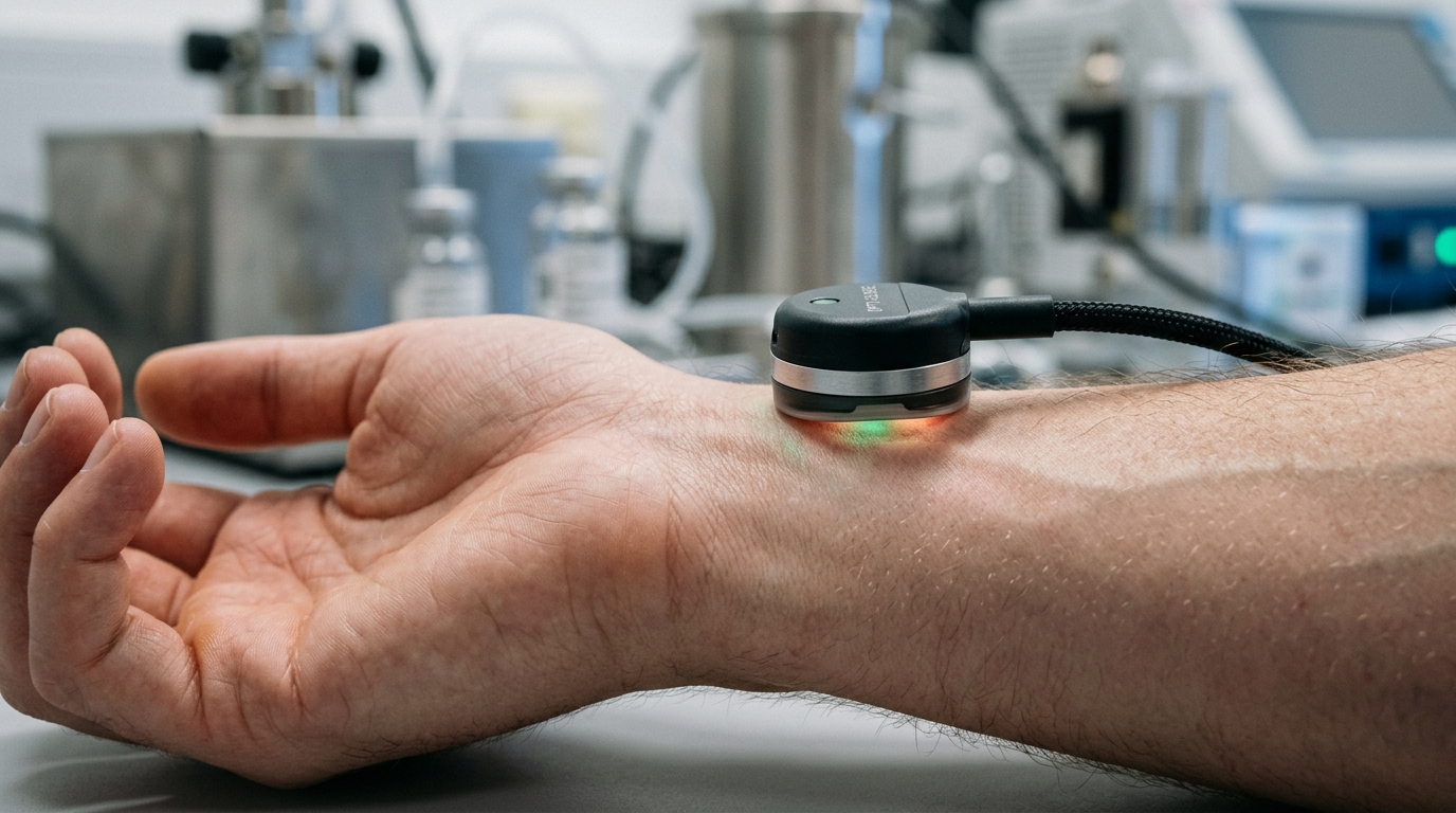
Despite the practical appeal of photoplethysmography, which is routinely collected via accessible finger-clip sensors, translating optical waveforms into arterial blood pressure curves constitutes an ill-posed inverse problem. Photoplethysmography measures pulsatile blood volume fluctuations across peripheral microvascular tissue rather than direct arterial lumen pressure. While both signals share timing benchmarks and gross morphological contours under controlled conditions, their biological relationship is fundamentally indirect, non-linear, and physiologically non-unique. The transmission of light through peripheral vascular beds depends on dynamic vascular compliance, local tissue perfusion, baseline vasomotor tone, ambient and body temperatures, sensor contact pressure, hardware-specific optical properties, autonomic regulation, and patient posture. Consequently, identical optical pulse morphologies can correspond to markedly different arterial blood pressure waveforms across different individuals or within the same patient as physiological states fluctuate. Simple optical waveforms cannot deterministically dictate arterial pressure contours without accounting for these confounding vascular mechanisms.
To train predictive models, researchers depend on public biobanks containing simultaneous recordings of invasive arterial lines and photoplethysmography. The dominant source in the literature is the MIMIC database family, particularly the MIMIC-III Waveform database, which captures physiological signals from roughly 30,000 intensive care patients at 125 Hz. However, raw clinical recordings suffer from extensive corruption, leaving only five to ten percent of each signal usable. Furthermore, lacking consistent subject identifiers in the broader MIMIC-III repository frequently forces investigators to use the smaller Matched Subset or risks mixing segments from the same patient across splits. An updated MIMIC-IV Waveform collection provides 198 patient records, with a 10,000-record release planned. Alternatively, the VitalDB repository records intraoperative signals from non-cardiac surgeries at 500 Hz, preserving comprehensive device metadata. Curated derivatives, such as the UCI Cuff-Less dataset and PulseDB, offer segmented files, but aggressive filtering in PulseDB—discarding pairs with correlation coefficients below 0.9—creates benchmarks that cannot be deployed without access to the invasive target.
Signal preprocessing and quality control represent pivotal stages governing downstream model accuracy. Across reviewed studies, signal validation strategies cluster into three distinct methodologies: rule-based template matching, classical feature extraction with machine learning, and deep neural networks. Template-based systems identify representative pulsations using running averages, adjacent pulse comparisons, or principal component analysis, discarding corrupted pulses through cross-correlation thresholds, dynamic time warping, or skewness signal quality indices classifying segments as excellent when skewness exceeds zero. Machine learning techniques extract curated temporal and spectral features to train support vector machines, C-SVC classifiers, or random forests, though comparisons show no consensus on superior performance. Meanwhile, deep learning approaches utilize one-dimensional convolutional neural networks, recurrence plot backbones, or autoencoders for anomaly detection. In pipeline preparation, cross-correlation serves as the primary alignment tool, correcting transmission delays reaching 500 milliseconds in raw records. Band-pass filtering commonly applies Butterworth architectures within a 0.1 to 8 Hz passband, while normalization requires local scaling because optical signals are recorded in arbitrary units.
Deep learning architectures overwhelmingly dominate research in continuous waveform prediction, with sequence-to-sequence U-Net variants representing the most prevalent model family. Originally designed for biomedical image segmentation, U-Net’s multi-scale paths and skip connections transfer effectively to one-dimensional signals by preserving fine temporal resolution. Following foundational work by Athaya and Tasbiraha utilizing vanilla U-Net frameworks on 256-sample windows across 100 patients, subsequent studies introduced architectural refinements. Hill and colleagues incorporated discrete systolic and diastolic values into inputs, used convolutional networks for quality assessment, targeted residual differences between optical and pressure contours, and validated models across patient splits. Other variations incorporate advanced blocks, including Vardhan’s Average Ensemble Block with self-supervised pretraining, Tang’s dual-stage W-Net with composite loss functions, and MultiResidual connections explored by Ibtehaz and Zhou. Mahmud and co-workers implemented a two-stage paradigm predicting discrete pressure values before estimating waveforms through U-Net++ augmented with attention-guided and bi-convolutional recurrent modules, while Ma and colleagues integrated adversarial ResNet-18 classifiers to enforce category independence prior to waveform reconstruction.
Beyond convolutional encoder-decoder frameworks, investigators have explored attention-based architectures, recurrent networks, and autoencoders to capture temporal dependencies. Aguirre and colleagues developed a bidirectional gated recurrent unit model with attention, integrating patient age and sex alongside first-derivative signals to predict averaged arterial pulsations. Notably, under rigorous patient-independent evaluation, their model demonstrated substantial mean absolute errors of 12.08 mmHg for systolic and 5.56 mmHg for diastolic pressures, illustrating how realistic testing tempers optimistic claims. Addressing artifact corruption, Bian and associates developed a Latent Space Constraint Transformer combining Swin-Transformer blocks, quantized codebooks, and spectral enhancers capable of reconstructing waveforms despite 10 to 90 percent input masking. In the autoencoder domain, Harfiya and colleagues established the largest evaluated cohort in the literature, training a long short-term memory autoencoder across 5,289 patients by freezing a pretrained optical encoder and fine-tuning an arterial reconstruction decoder. Conversely, alternative paradigms like unpaired CycleGAN architectures required patient-specific model training, sacrificing generalizability across unseen individuals.
A critical distinction separates experimental optical algorithms from commercial monitors outputting non-invasive arterial pressure waveforms. Currently, no commercial technology reconstructs continuous arterial blood pressure waveforms solely from a passive, clip-on pulse oximetry probe. Instead, commercial waveform monitors rely on active physiological manipulation or tonometry. Volume-clamp devices, including Finapres, Nexfin, and CNSystems platforms, implement the method introduced by Jan Peňáz in 1973. These systems use an inflatable finger cuff paired with an infrared sensor within an active feedback loop, continuously adjusting external pressure to maintain zero transmural pressure across the digital artery, compensated by Wesseling’s Physiocal algorithm tracking vascular tone. Alternative solutions, like the CareTaker monitor, apply low sub-diastolic cuff pressure and analyze pulse derivatives using piezoelectric sensors and pulse decomposition algorithms, though raw outputs remain restricted to arbitrary units. Radial applanation tonometry instruments like HealthSTATS BPro and transfer-function systems like SphygmoCor XCEL similarly require external brachial cuff calibration to scale continuous arterial contours into absolute pressure units.
The absence of clip-on optical waveform monitors in commercial healthcare is compounded by an acute regulatory and standardization deficit. Although existing commercial non-invasive devices possess United States Food and Drug Administration 510(k) clearances, clearance indicates only substantial equivalence to historical predicate devices rather than direct validation of continuous waveform morphology. The governing clinical validation standards—specifically ISO 81060-2, IEC 80601-2-30, and the historical AAMI SP-10 protocol—establish strict performance requirements exclusively for discrete systolic and diastolic pressure determinations from automated or manual sphygmomanometers. None of these regulatory benchmarks provide validation protocols, acceptable error thresholds, or morphological standards for continuous arterial pressure waveforms compared against in vivo arterial lines. Consequently, regulatory clearance cannot be construed as evidence that a device produces physiologically valid waveform shapes. This regulatory void leaves clinical engineers and downstream analytical algorithms without clear metrics to judge whether non-invasive waveform outputs reliably reflect cardiovascular dynamics.
Methodological shortcomings within the academic literature further obscure algorithmic capabilities. A pervasive vulnerability involves omitting subject-based partitioning; splitting datasets by random segments allows networks to memorize individual baseline pressures, generating artificially low error rates that collapse on unseen patients. Furthermore, optimizing models using mean squared error produces smoothed waveforms that obscure the systolic upstroke and dicrotic notch, while isolated loss penalties on worst-fitted samples waste representational capacity. These concerns are reinforced by empirical trials, including the Microsoft Aurora-BP study, where optical features provided only marginal improvements over baseline calibration across differing postures and environments. Cardiovascular experts note that optical pulse analysis has yet to convincingly demonstrate blood pressure measurement even in ideal conditions.
Subject of Research: Machine learning methods for estimating continuous arterial blood pressure waveforms from photoplethysmography
Article Title: A narrative review of methods for estimating blood pressure waveforms from photoplethysmography
Article References: Olszewski, B., Mataczynski, C., Ostrowski, Z., & Kasprowicz, M. (2026). A narrative review of methods for estimating blood pressure waveforms from photoplethysmography. Journal of Clinical Monitoring and Computing. https://doi.org/10.1007/s10877-026-01521-0
Image Credits: AI Generated
DOI: 10.1007/s10877-026-01521-0
Keywords: photoplethysmography, arterial blood pressure, waveform reconstruction, continuous hemodynamic monitoring, deep learning, U-Net, volume-clamp method, MIMIC-III, VitalDB, signal quality assessment, pulse contour analysis, dicrotic notch, subject-based split, FDA 510(k), ISO 81060-2, transformers, autoencoders
News Source: Ophelia Keating. (November 17, 2026). Optical Blood Pressure Waveform Reconstruction Stalls Behind Validation Gaps and Physiological Limits. Scienmag.